

We know that viruses infect cells, but what does that process actually look like? Sure, diagrams can be helpful, but there’s nothing quite like seeing the real thing. Trouble is, doing so with the kind of microscopes you get in a classroom can be pretty difficult – but in some first-of-its-kind footage, researchers have successfully risen to the challenge.
The rest of this article is behind a paywall. Please sign in or subscribe to access the full content.The team, led by Professor Masaharu Takemura at the Tokyo University of Science, did so by making use of a particular type of virus: Mimivirus.
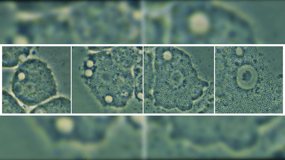
The vast majority of viruses are far too small to be seen with the standard light microscopes we use to learn about cells in school. Mimivirus, however, is a giant virus, with a total diameter of around 750 nanometers (or 0.00075 millimeters) – not big enough to see with the naked eye, but more than big enough for a light microscope.
To attempt to visualize how it infects cells in real time, the researchers had to put it in the presence of a target. In this case, that target was Acanthamoeba castellanii; this species of amoeba and others in its genus are prevalent in the environment (and can occasionally cause us issues).
Like viruses, amoebae aren’t exactly the easiest to visualize – not because of size, but because they’re always on the move if you put them in a liquid. To combat that, the team grew A. castellanii in agar, a thick, jelly-like substance that’s a staple of the microbiology toolkit.
This turned out to be the perfect recipe for successfully using a light microscope to capture the infection of A. castellanii by mimiviruses in real time.

"For the first time in the world, we have succeeded in continuously visualizing the events that are believed to occur in viral infection over a long period of time – such as the proliferation of the virus, its release from cells, and the death of cells during the process,” said Takemura in a statement.
The researchers now hope that the footage they captured can be used in the classroom, both in schools and colleges. While the goal is to show the film rather than get the students to carry out the experiment themselves, it’s thought that it could give them a better understanding of the infection process and a broader perspective on virology as a whole.
It has the potential, Takemura concluded, to enhance “students' understanding of virus proliferation mechanisms and [highlight] the biological significance of viruses, their impact on host cell fate, and their role in ecosystems."
The study is published in the Journal of Microbiology & Biology Education.