Health and Medicine
 PUBLISHED





Capturing a seizure as it happens in the brain is about as easy as trying to hold water between your hands. The electrical patterns move so quickly that getting a good-quality 3D image has been next to impossible with existing methods.
Enter a team at the University of Georgia and their new imaging system that has captured a seizure in a zebrafish brain in extraordinary, high-resolution 3D.
I mean, just watch this.
.gif "gif showing a zebrafish brain rotating with areas of orange lighting up and moving through it to represent seizure activity")
“We developed a light-sheet microscope that allows rapid volumetric imaging with real-time correction of aberrations – imperfections in the way a microscope forms an image,” said team lead Peter Kner in a statement.
“Most imaging of seizure events in zebrafish have only captured 2D images, but our system allows 3D high-resolution imaging over a larger volume than was previously possible.”
Seizures: what they are and how they happen
Seizures are most often associated with a diagnosis of epilepsy. But there are lots of different types of seizures, and anyone could experience an isolated seizure at some point in their life without being diagnosed with a chronic condition.
With this study, we’re talking about epileptic seizures, which arise when the brain’s electrical signaling is disrupted or too many signals are being sent at once.
Epileptic seizures can be broadly divided into two categories: focal and generalized. Focal seizures affect just one part of the brain (although it can be a large part), whereas generalized seizures appear in both hemispheres at once.
Not every seizure starts in the brain. Functional seizures, for example, can resemble epileptic seizures outwardly, but they aren't associated with unusual activity in the brain and are instead caused by an extremely traumatic or stressful experience.
Not all epileptic seizures cause convulsions or loss of consciousness, either – popular media tends to focus only on this type, called tonic-clonic seizures, but there’s a huge diversity of experiences.
Illustrator Helena Napier shared her experience of living with temporal lobe epilepsy (a common form of focal epilepsy) with the Epilepsy Society, describing her first memory of a seizure during a church service at the age of 10:
“It started off extremely pleasant, like deja-vu, rising from my stomach to my throat. Then the stench of petrol entered my nostrils and the altar ahead gave way to a different scene.”
“I ‘saw’ what I perceived to be an ocean of bones, each wrapped in its own individual green leaf. Hundreds of them swam together calmly under a stretch of clear blue sky. The pew my small fingers clung to dissolved, and I was plunged simultaneously into a sense of both total disorientation and incredible clarity.”
A microscope that's seven times faster
The goal of the new study was to gain a better understanding of how seizure activity moves through the brain and how it’s influenced by a gene called gad1b.
The team used the neuroscientist’s favorite model organism, the zebrafish. Having previously imaged seizure activity in the zebrafish brain in 2D, they wanted to progress to 3D.
They used light-sheet microscopy, which employs a thin sheet of light that illuminates the subject from one side and “is a powerful tool for imaging live organisms,” the team writes in their paper.
Using a thin band of light limits damage to the Zebrafish that can be caused by excessive light exposure in other types of microscopy.
Previously, the researchers had developed a light-sheet system that could capture an image in 1.75 seconds, but even that isn’t fast enough to follow the evolution of a seizure.
After a few improvements, they managed to up this speed by seven times, capturing 600 continuous 3D images in just 2.5 minutes. They tested the system in seven zebrafish larvae.
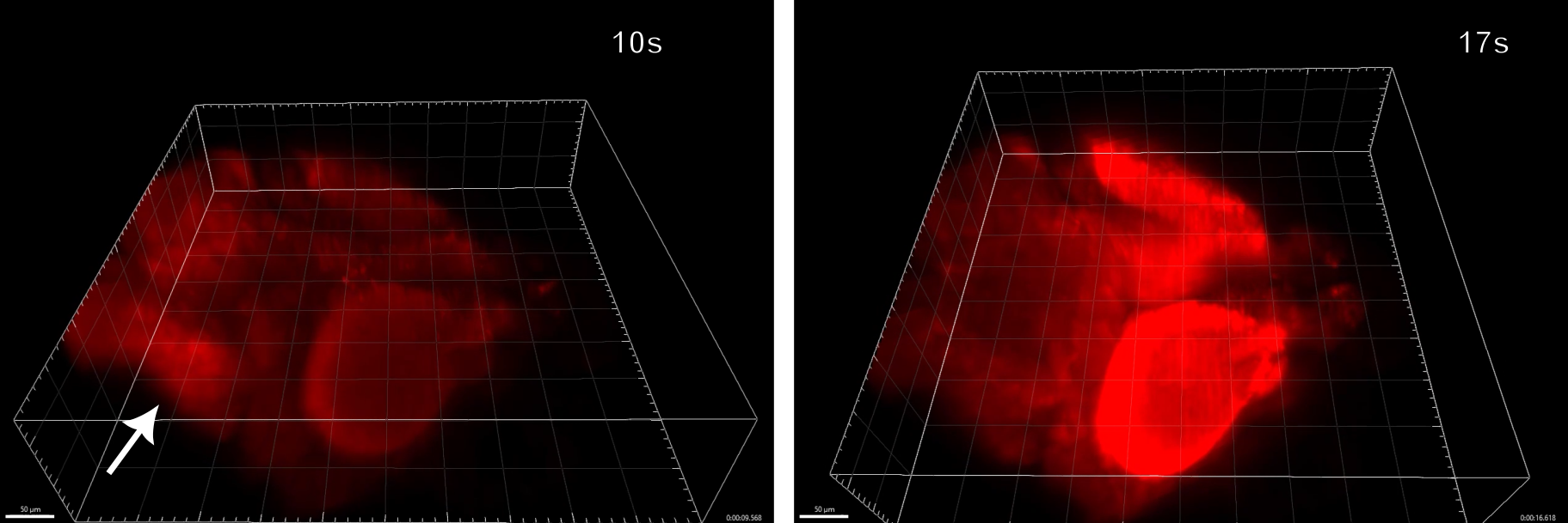
Watching the footage, they could see that seizure activity in the zebrafish started towards the back of the brain and spread forwards, lasting for tens of seconds in total. Some previous studies agree with this, but others don't, so there's more to be done to understand whether this is the definitive mechanism for seizures in zebrafish.
“The detailed imaging information available from our fast volumetric imaging technique could provide new insights into the mechanisms of seizure formation and propagation, helping guide the development of more effective therapies,” said Kner.
The team now wants to repeat the feat with zebrafish lacking the gad1b gene to compare their seizure patterns. Looking ahead, they hope a more detailed understanding of how seizures work will positively impact epilepsy treatment in humans.
“More generally, the approach could improve our understanding of how the brain operates, helping inform the treatment of various brain diseases and disorders.”
The study is published in the journal Biomedical Optics Express.