

It was announced earlier this month that Eric Betzig of the Howard Hughes Medical Institute (HHMI) in Ashburn, Virginia will be sharing the 2014 Nobel Prize in Chemistry as a reflection of his contribution to "the development of super-resolved fluorescence microscopy.” He is continuing to advance the field of microscopy by leading a team that developed a lattice light-shield microscope which offers high-resolution, 3D images of moving, living cells and organisms. The paper describing the microscope has been published in Science.
Imaging three-dimensional movement of small structures in living cells is a tricky balancing act. If the images are high resolution, the images can’t be collected quickly, resulting in a discontinuous series. The powerful beam of light used to pick up the fluorescent markers is very harsh and damages the cells, interfering with the study of the structures.
Betzel’s team attempted to resolve this problem by changing the way the sample is illuminated. They developed the Bessel beam plan illumination microscope in 2011 that uses a sheet of light, rather than a single powerful beam. Dispersing the light across a plane is much easier on the cells and allows for high-res images to be collected quickly. Though this approach is gentler, the beam’s shape causes the sides of the sample to appear slightly out of focus—a problem they have been trying to resolve for a long time.
Movie 11 High Resolution from HHMI NEWS on Vimeo.
Now, they believe they have fixed the resolution problems by combining features from the Bessel beam and traditional single beams of light. Rather than a single plane of light illuminating the subject from the side, seven parallel beams have been arranged to form a planar lattice pattern. There is some interference between the beams, but they can be arranged to provide light where it is needed to sharpen those blurry edges. This arrangement is also much easier for the cells, which is very important for studying these structures.
"What was shocking to us was that by spreading the energy out across seven beams instead of one, the phototoxicity went way down," Betzig said in a press release. "What I learned from that experience is that while the total dose of light you put on the cell is important, what's far more important is the instantaneous power that you put on the cell."
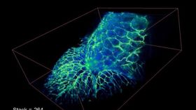
This technique can be used to image proteins as they travel in an out of cells, which allows researchers to see cells, tissues, and small organisms as they are. This brings a deeper level of understanding of the structure’s purpose. This has a wide range of applications, including some that the researchers claim have not even been thought of yet.
In addition to the one used in Betzig’s lab, a second microscope has been built and donated to HHMI’s Advanced Imaging Center. This microscope is available to use, completely free of charge. Anyone interested in using this technology are welcome to submit proposals.
Movie 9 High Resolution from HHMI NEWS on Vimeo.
If you’re interested in seeing more videos using this technology, check out the lab’s Vimeo channel for the complete video collection.