

New research published in the journal Science Advances describes a new process that enables scientists to look deep into organs and nervous systems of animals by turning their external tissues translucent. The ghostly see-through creatures, examples include squid, worms, fish, and salamanders, give a stunning (if not a little freaky) insight into the complex internal workings of animals.
The rest of this article is behind a paywall. Please sign in or subscribe to access the full content.The best practice for examining cells and organ structures used to involve cutting a specimen’s tissues into thin layers and peeling each apart to get a better look before using the information gleaned from the organ wafers to build a 3D model of the functional structure. The process is unfortunately every bit as tedious as it sounds, being both time-intensive and prone to failure.
Researchers decided it would make their lives easier creating a technique that essentially erases the surrounding tissues by making them transparent. This process enabled them to isolate complex structures from organs such as the brain without painstakingly carving up such a delicate organ.
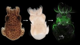
Getting the right level of transparency however didn’t prove easy as pigmented tissues were less likely to become sufficiently clear in order to be able to see anything. A new method developed by a team of researchers from a number of collaborating institutions in Vienna combines tissue clearing with pigment removal in a sophisticated and effective process dubbed “DEEP-Clear”.

A key observation in the breakthrough was that by using different chemical treatments the researchers could speed up the process of tissue clearing, preserving the integrity of the delicate specimens. The DEEP-Clear was also able to detect a variety of important biomolecules for observation, which meant specific proteins, DNA markers, and RNA could be observed through the transparent tissues of intact specimens.
They took their DEEP-Clear imaging one step further in combining their findings with the latest generation of light-sheet microscopes, which use two-dimensional laser light to scan a sample. The scan creates a 3D model on a computer that can then be printed to create a to-scale model of the observed structures and organs.
"This versatility of DEEP-Clear makes it a highly attractive tool to explore a range of animals for which standard tissue clearing techniques currently would not be sufficient," explained researcher Karim Vadiwala from Max Perutz Labs, Vienna, in a statement.
It’s hoped the technology can be harnessed to uncover clues to hard-to-analyze specimens such as worms, fish, and salamanders that are known to regenerate parts of their central nervous system. That humans don’t have this ability indicates we lost certain molecular processes along the long road of evolution and understanding what these are could further our ability to help those with disabilities such as paralysis from traumatic accidents.
"Visualizing the responsible stem cells, and investigating their molecular make-up, or their contribution to regenerated tissue, will be greatly facilitated by DEEP-Clear," said Dr. Florian Raible from Max Perutz Labs, Vienna, who coordinated the study.