

The 12th Nikon Small World in Motion competition saw hundreds of entries from all over the world. A sister to the Nikon Small World Photomicrography Competition, both reward skill and excellence in video and photography under the microscope.
The rest of this article is behind a paywall. Please sign in or subscribe to access the full content.The video portion encompasses any movie or digital time-lapse photography taken through the microscope, opening up new ideas to the audience and, of course, impressing the panel of judges. The entries are judged on originality, technical skill, informational content, and visual impact.
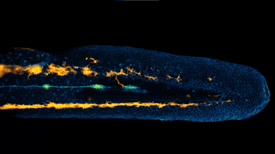
This year's winner is Dr Eduardo E. Zattara for his incredible time-lapse of neural crest cells migrating in a zebrafish embryo, which you can check out below. Highlighting the dynamic study of evolutionary developmental biology, sensory organ progenitors (colored in green) migrate along the lateral line of the zebrafish embryo. The orange cells, melanocytes, also move to reach their final positions below the zebrafish’s skin.
“This recording came out very clean and required almost no post-processing. It is an astonishing display of the dynamics of neural crest cell migration,” said Zattara in a statement seen by IFLScience.
“The result was a video that was both biologically informative and visually striking."
Second place was awarded to Dr Christophe Letterier for his 12-hour time-lapse video of monkey cells. In the video below, you can see the plasma membrane labeled in orange and the DNA colored blue. The challenge here for Letterier, was to keep the cells alive for the time frame of the video, with closely controlled temperature and humidity as well as minimal phototoxicity from laser illumination.
Dr Ahmet Karabulut was awarded third place for his video (below) of sea anemone neurons and stinging cells showing their dynamic processes.
In total, five top prizes were awarded to the most impressive videos, with several others taking honorable mentions and $100 each. See the full gallery of award winners here.