

Organoids modeled on the structure of the human spinal cord have been shown to match the behavior of an injured spine. Now, a treatment that has worked well in mice has been applied to the organoids, and it proved highly successful.
The rest of this article is behind a paywall. Please sign in or subscribe to access the full content.Research on many of the body’s parts has been fast-tracked using organoids: small, simplified versions of organs grown from human stem cells, but some organoids are more faithful replications than others. Scientists at Northwestern University have called their lab-grown nerves “the most advanced organoid model for human spinal cord injury to date.”
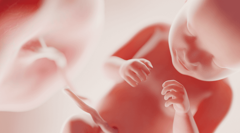
The spine is a wonder of evolution, but the fact that human versions lack the rejuvenation powers of some lizards and amphibians is a deep disappointment. Injuries cause cell death, inflammation, and produce a mass of scar tissue known as glial scarring. The scarring both physically prevents the nerves from regenerating and changes the chemistry to do the same. Although techniques to circumvent the damage have given patients renewed mobility, they are far from a dream solution.
Organoids that accurately reflect this damage provide a way to test treatments faster than using animal models and without the same ethical concerns. A sufficiently advanced organoid would also be a more accurate representation of the human spine than rodents separated from us by tens of millions of years of evolution.

Demand for spinal therapy that works is so high that many approaches are under investigation, including “dancing molecules,” which form a gel around a wound site and signal cells to start recovery. The gel then breaks down into nutrients that the cells can absorb. Dancing molecules rapidly reversed paralysis in mice with damaged spinal cords in one study, which was particularly impressive since it required only one injection.
A demonstration of organoid recovery after dancing molecule treatment has bolstered hopes that even if people suffering paralysis may not soon be dancing themselves, they may regain significant function.
“One of the most exciting aspects of organoids is that we can use them to test new therapies in human tissue,” dancing molecules' inventor, Northwestern’s Professor Samuel Stupp, said in a statement. “Short of a clinical trial, it’s the only way you can achieve this objective. We decided to develop two different injury models in a human spinal cord organoid and test our therapy to see if the results resembled what we previously saw in the animal model.”
The team are excited by the results. “After applying our therapy, the glial scar faded significantly to become barely detectable, and we saw neurites growing, resembling the axon regeneration we saw in animals,” Stupp continued. “This is validation that our therapy has a good chance of working in humans.”
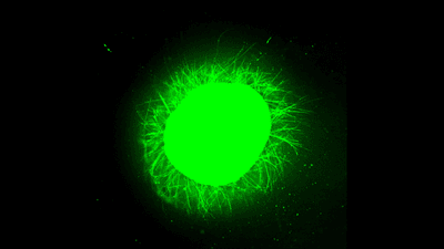
 compared to one treated with slow-moving molecules (right) using otherwise similar chemicals.")
Stupp’s team are not the first to make spine organoids, but they claim theirs are far larger and more accurate, providing the first useful lab-grown model for testing therapies like dancing molecules. Among other advances, the Northwestern organoids are claimed to be the first to include microglia, the central nervous system’s immune cells.
Since inflammatory responses to traumatic spinal cord injuries cause the scarring that presents one of the major obstacles to regeneration, microglia are essential for a full picture. “It means that our organoid has all the chemicals that the resident immune system produces in response to an injury. That makes it a more realistic, accurate model of spinal cord injury,” Stupp said.
By patiently growing the organoids over four months, the team coaxed the organoids to about 3 millimeters (0.12 inches) wide, which they say is large enough to reflect injuries with some accuracy.
The name of Stupp’s treatment comes from the fact that the gels are made of nanofibers that are made to move together, or “dance”, under the creators’ control.
“Given that cells themselves and their receptors are in constant motion, you can imagine that molecules moving more rapidly would encounter these receptors more often,” Stupp said in 2021. “If the molecules are sluggish and not as ‘social,’ they may never come into contact with the cells.”
Stupp’s past success with mice involved injecting the dancing molecules 24 hours after the spinal injury. In the organoids, the team caused two sorts of damage, mimicking the injuries from a knife and a car accident, respectively. The scar tissue reflected what occurs in a real spine.

The team tracked the cell response and the chemicals produced. When the dancing molecules were introduced, they quickly formed a scaffold around the cells. Neurons, and their projections, neurites, were not only stimulated to grow, but did so in ordered patterns, rather than spreading haphazardly.
In control experiments, molecules that didn’t participate in this safety dance proved no friends of the neurites, which were left behind.
The hope is that the fresh neurites will restore the connections whose severing causes paralysis.
If clinical trials are planned, the team are keeping them close to their chest, instead talking about working on more advanced organoids, for example, by including blood vessels. In particular, they want to make organoids that represent old injuries. This would make the work relevant to people with existing damage, rather than the small portion of the 250,000-500,000 estimated to suffer new spinal injuries each year, who could be treated soon after the damage occurs.
Stupp and co-authors don’t think their work’s potential stops there. “Similar models could also be extended to investigate the problem of traumatic brain injury,” they write.
The study is published in Nature Biochemical Engineering.