

Researchers have discovered 31 new species in the South Atlantic Ocean during a two-week expedition – a feat made possible by new imaging technology that let them study some of the ocean's most fragile animals without bringing them to the surface.
The rest of this article is behind a paywall. Please sign in or subscribe to access the full content.The Designing the Future 3 expedition took place aboard the Schmidt Ocean Institute's research vessel Falkor (too) off the coast of Fortaleza, Brazil, between April 15 and 30. It's the third in a series of expeditions aiming to demonstrate new and better ways of documenting the extraordinary diversity of life in the ocean.
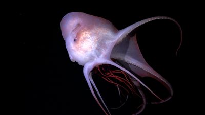
The new species, ranging from jellyfish to amphipods, were all found in the Earth's largest and least explored habitable ecosystem known as the midwater. This is the layer of water between the ocean's sunlit surface and the seafloor.
 uses a custom-built camera array to photograph midwater creatures")
"The largest habitat on Earth, the midwater, is filled with incredible animals we are only just starting to understand," said Dr Karen Osborn, the expedition's chief scientist from the Smithsonian National Museum of Natural History, in a statement.
"I continue to be fascinated by the fantastic variety of solutions they have evolved to survive in this formidable environment, and that drives me to keep asking questions about our ocean."
For Dr Kakani Katija at Monterey Bay Aquarium Research Institute's Bioinspiration Lab, that sense of the unknown is what makes midwater expeditions so compelling. "You could go to almost any place – near-shore or far offshore – and come across a midwater animal that's unknown to science," she told IFLScience.
You're constantly rolling with the situation – you have very little control over what's happening.
Dr Kakani Katija
Many of these animals are soft and gelatinous, which makes them notoriously difficult to study. Conventional methods involve catching them in nets and pickling them in jars, but that leaves the animals looking nothing like they did in the water. Because of this, it can take scientists decades to identify and describe a new species, said Katija.
But by using new technology that lets researchers visualize the structure and biomechanics of these delicate deep-sea animals while they are still in their natural habitat, the Designing the Future 3 team was able to cut this down to just a few days.
The systems – DeepPIV (particle image velocimetry) and EyeRIS (remote imaging system) – were developed by the Bioinspiration Lab and mounted on the expedition's remotely operated vehicle, SuBastian. Both are non-invasive tools that use lasers to scan organisms and build detailed 3D models.
DeepPIV uses a sheet of laser light to create 3D scans of the internal structures of translucent soft-bodied organisms, while EyeRIS can capture the external structure of both translucent and opaque creatures.
SuBastian was also equipped with a collection jar for gathering tissue samples for DNA sequencing and a Shadowgraph camera that can image finer details not visible in the 3D scans.
The new species identified with the new technology includes an amphipod, a gossamer worm, nine jellyfish, seven siphonophores, seven comb jellies, four larvaceans and two giant rhizarians. In a statement, the team wrote that they observed far more diversity in the midwater than they had expected.
")
Among the most striking finds were the seven new siphonophores. These are relatives of jellyfish and corals that are made up of independent units called zooids. Each zooid develops from a single fertilized egg and so, in a sense, each is an individual animal. But they must combine together to form functional colonies that are able to digest food, reproduce, and move around using jet propulsion.
"Our organs are interior to us – we have a liver, a spleen, and they're all contained within our skin, so we're considered one organism," said Katija. "What's interesting with siphonophores is that they're considered colonies, but they have the equivalent of organs: units called zooids, which have evolved very specific functions in feeding, reproduction, or defense."
The team also used Squid, an open-source microscope developed at Stanford, to carry out the first 3D imaging at sea of an organism's internal cellular structures. One of the animals imaged was a large single-celled microbe called a protist, and researchers were able to observe how its cellular structure interacted with its glass skeleton.

The work on board Falkor (too) was varied and constantly changing, said Katija. "That's what makes expeditions so interesting, challenging, and all-involving," she said. "You're constantly rolling with the situation – you have very little control over what's happening. You can lay out a plan for every hour, but once you're offshore, those plans more often than not go out the window. That was very true with this one."
The expedition collaborated with the FathomNet project. This aims to build a library of labeled imagery to train AI to identify marine life through recruiting users to its mobile app FathomVerse. For the first time, the app featured contributions from an expedition still underway, letting anyone, anywhere, take part in the discoveries as they happened. Footage from SuBastian's cameras was also livestreamed to viewers online.