

A new, easy-to-use, and open-source platform is streamlining the process of segmenting large images for scientists. Available through a web browser without the need for arduous configuration and software installation, it can be used for all sorts of 3D imaging and marks a significant improvement on conventional approaches to segmenting large images.
Biomedisa, a Biomedical Image Segmentation App, is free to use and hopes to open up this valuable arm of investigation to scientists who lack the extensive computer-know-how required for existing image analysis. The end result is stunning images that reveal in great detail the constituent parts of complex organisms and structures, big or small. The team behind the platform presented it in a paper in Nature Communications.
Imaging is a vital tool in many scientific disciplines, from looking at brains to revealing incredible details about ancient Egyptian mummified cats. To create them, data is obtained from biological imaging tools such as X-ray computed tomography (CT), magnetic resonance imaging (MRI), and optical microscopy. However, once these images have been obtained, they usually require a complex and time-consuming process of isolating individual structures from the overall structure by endlessly segmenting the images, sometimes slice by slice. This frustrating process has often rendered 3D imaging an impractical tool in time-sensitive investigations in medicine.

Historic practices have been highly effective in sucking the fun out of scientific discovery, so it will no doubt be an enormous relief for many practicing researchers that image analysis has at last been streamlined. In recent years, the leaps and bounds made in imaging technologies have increased the demand for efficient 3D modeling, which was used to demonstrate the lung damage caused by the novel SARS-CoV-2 coronavirus. Bottlenecking a labor-intensive and error-prone task such as 3D image analysis on such research can delay progress and lead to false conclusions, so this new, open-source technology will support both researchers and scientific progress across many disciplines.
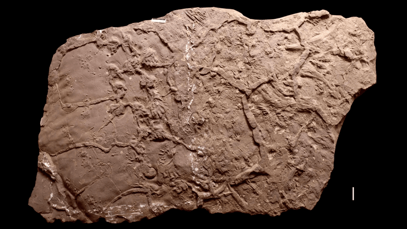
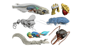
The new platform’s recent paper includes a Medaka fish with segmented skeleton and selected internal organs, a mouse’s molar tooth showing the enamel and dentine, and a human heart with segmented heart muscle and blood vessels. It’s also been used to segment a fossil specimen of a parasitoid wasp from Baltic amber as well as the tracheal system of a hissing cockroach and a claw from a theropod dinosaur from Burmese amber. The star of the piece however is a Trigonopterus weevil, and if you’ve ever wanted to see such an animal collapse into its constituent parts like a house of cards, well, today’s your lucky day.
“Our explicit aim was to create a freely available, user-friendly and widely applicable tool to improve the tedious manual segmentation procedure that still dominates 3D image analysis in many biomedical disciplines,” write the study authors. “Biomedisa was developed for CT and MRI but is generally suitable for many more types of volumetric image data, e.g. from confocal laser scanning microscopy, focused ion beam scanning electron microscopy or histological imaging. Even though the development of Biomedisa was motivated by applications from biology and medicine, it can also be employed for 3D data from other disciplines, e.g. geology, materials science, and non-destructive testing.”