


The microscopic world has never looked as beautiful as what has been captured by the winners and the honorable mentions of the second-ever Global Image of the Year Life Science Light Microscopy Award competition. Multinational corporation Olympus is behind this contest, which recognizes and celebrates life science photography. This is not just an important scientific endeavor to understand the world better, it has also incredible artistic value.
The competition has four winners: a global winner and three regional prizes covering Asia, the Americas, and Europe, Middle-East, and Africa (EMEA). The winners were selected from almost 700 submissions coming from 61 countries.
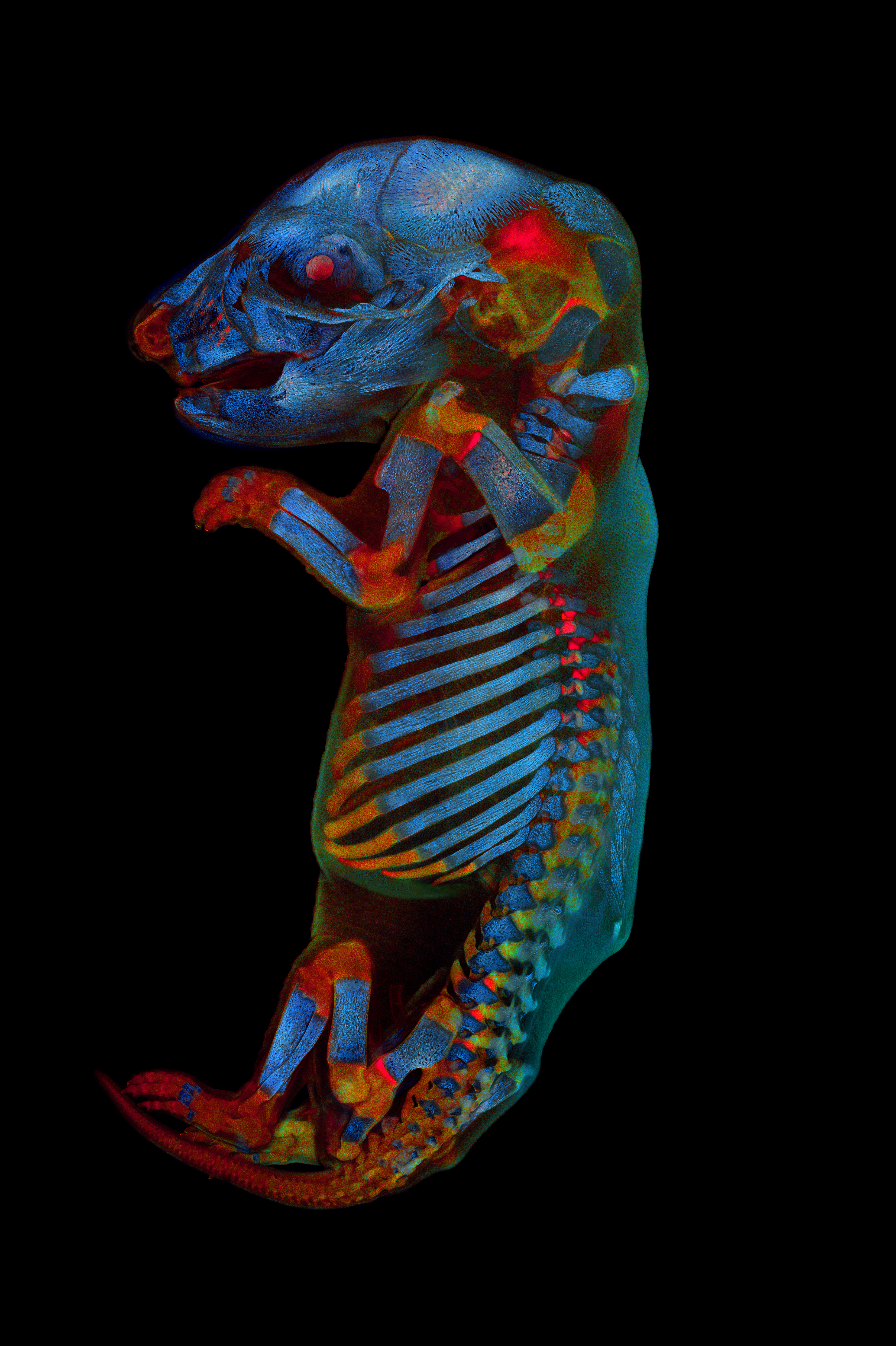
For this year, the global winner is Werner Zuschratter from Germany. He snapped a fantastic image of a whole rat embryo, captured using a confocal microscope. The regional winner for Asia was XinPei Zhang from China, who imaged the scales from the wings of over 40 species of butterflies one by one and then assembled them into this image.
The prize for the Americas goes to Justin Zoll from the US, who snapped a beautiful image of L-glutamine and beta-alanine crystals. Grigorii Timin (Switzerland) takes the prize for EMEA imaging the collagen fiber and dermal pigment cells from African house snake embryonic skin.

“Not only did we see a record number of submissions, but the quality and creativity of those images were exceptional. It’s amazing to see the unexpected art people capture with a microscope,” Satoshi Nakamura, Vice President of Scientific Solutions Global Marketing at Olympus Corporation, said in a statement.

The judges this year have also selected nine images that deserved honorable mentions. You can check them out on the Instagram carousel below:
"Our aim for the competition is to demonstrate both the artistic and scientific value of images captured under the microscope and further, to encourage people in every corner of the world to look at scientific images in a new way, appreciate their beauty, and share with each other,” Lee Wagstaff, Vice President, Life Science Sales and Marketing, Olympus Corporation of the Americas told IFLScience.