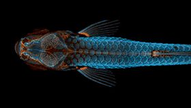


An image of an electric blue zebrafish with orange insides has won the top prize in the Nikon Small World Photomicrography Competition 2020.
This year marks the 64th Nikon Small World Photomicrography Competition, the world's oldest and most prestigious photo competition looking at the strange and beautiful world found beneath the microscope.
This year’s top prize was awarded to Daniel Castranova, assisted by Bakary Samasa while working in the lab of Dr Brant Weinstein at the US National Institutes of Health, for their image (above) of a juvenile zebrafish that had been fluorescently “tagged” to show its scales (in light blue) and its lymphatic system (orange) using confocal microscopy and image-stacking.
Not just a visually stunning image, it also shows the intriguing scientific discovery that this species of fish has lymphatic vessels inside their skull; a discovery that opens up the prospect of further research into treatments for diseases that affect the brain.
"Lymphatic vessels in the nervous system of mammals were first described in 2015, and since then major advances have been made in understanding how they function, and their role in disease," Castranova, who shot the winning image, told IFLScience. "Until now, it was unknown if fish had similar vessels. We are excited about this discovery because these vessels are much easier to image in zebrafish, making them a fantastic model to understand different aspects of the way these vessels grow and function."
Describing the winning photo, he added: "I chose to display the egfp [a green fluorescent protein] in orange and the mcherry [red fluorescent protein] in blue because I found the combination not only aesthetically pleasing, but it also makes the image more accessible to people with red-green color blindness."

Second place was awarded to Daniel Knop for his image of the embryonic development of a clownfish (Amphiprion percula), also known as the stripy orange fish that starred in the movie Finding Nemo. Using image-stacking, Knop showed development on days 1, 3 (on both the morning and evening), 5, and 9, detailing from the hours after fertilization to just a few hours before hatching.
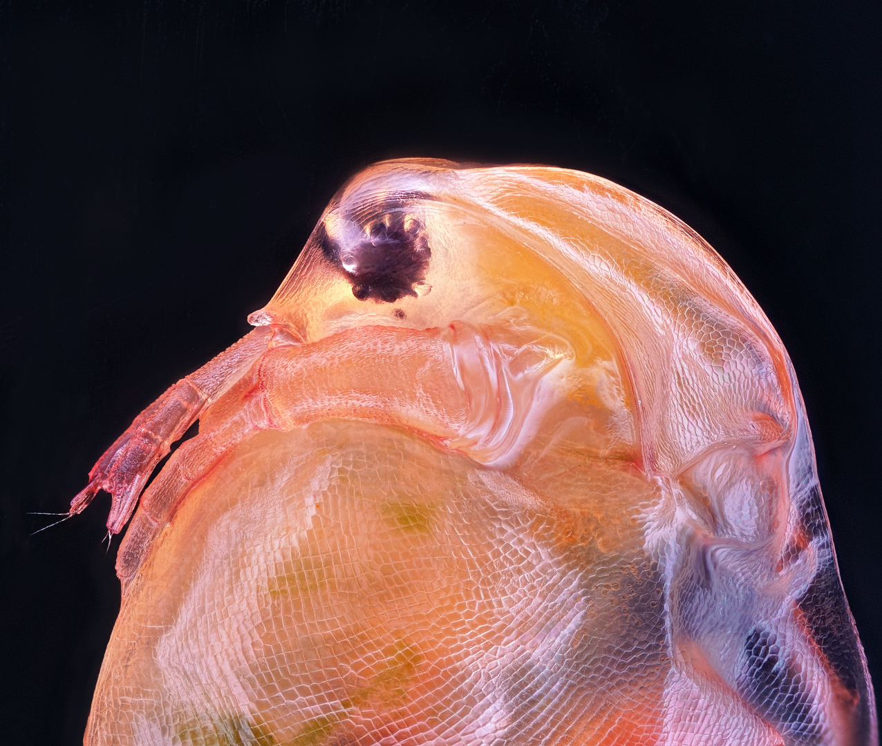
Third place was captured by Small World veteran Dr Igor Siwanowicz from the Howard Hughes Medical Institute in Virginia for his incredible picture of a “tongue” (technically known as a radula) belonging to a freshwater snail. Dr Siwanowicz also scooped second prize during last year's competition, the winners of which can be found here.
A number of the competition's winners and top picks can also be seen below.
If these stunning images tickle your fancy, then be sure to check out the Nikon Small World in Motion Competition, the sister video competition of the longstanding Nikon Small World still-photography competition.