Nature
 PUBLISHED



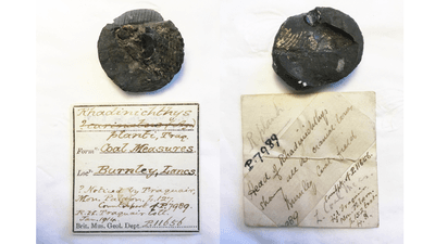


Fossils are typically bones, teeth, or mineralized versions of these hard tissues that act as a record of species past. While these are still rare and important findings, for one fish fossil, the landscape has managed an even more incredible feat: preserving the soft brain tissues of a 300-million-year-old animal in exquisite detail.
The minnow-sized fish fossil was discovered near the village of Trawden in Lancashire, UK. Known as Trawdenia planti, the fish passed the years since the Carboniferous period entombed in soapstone. Not only was the skull preserved, but the soft tissues inside the brain helped the researchers learn more about how these fossil fish were put together.
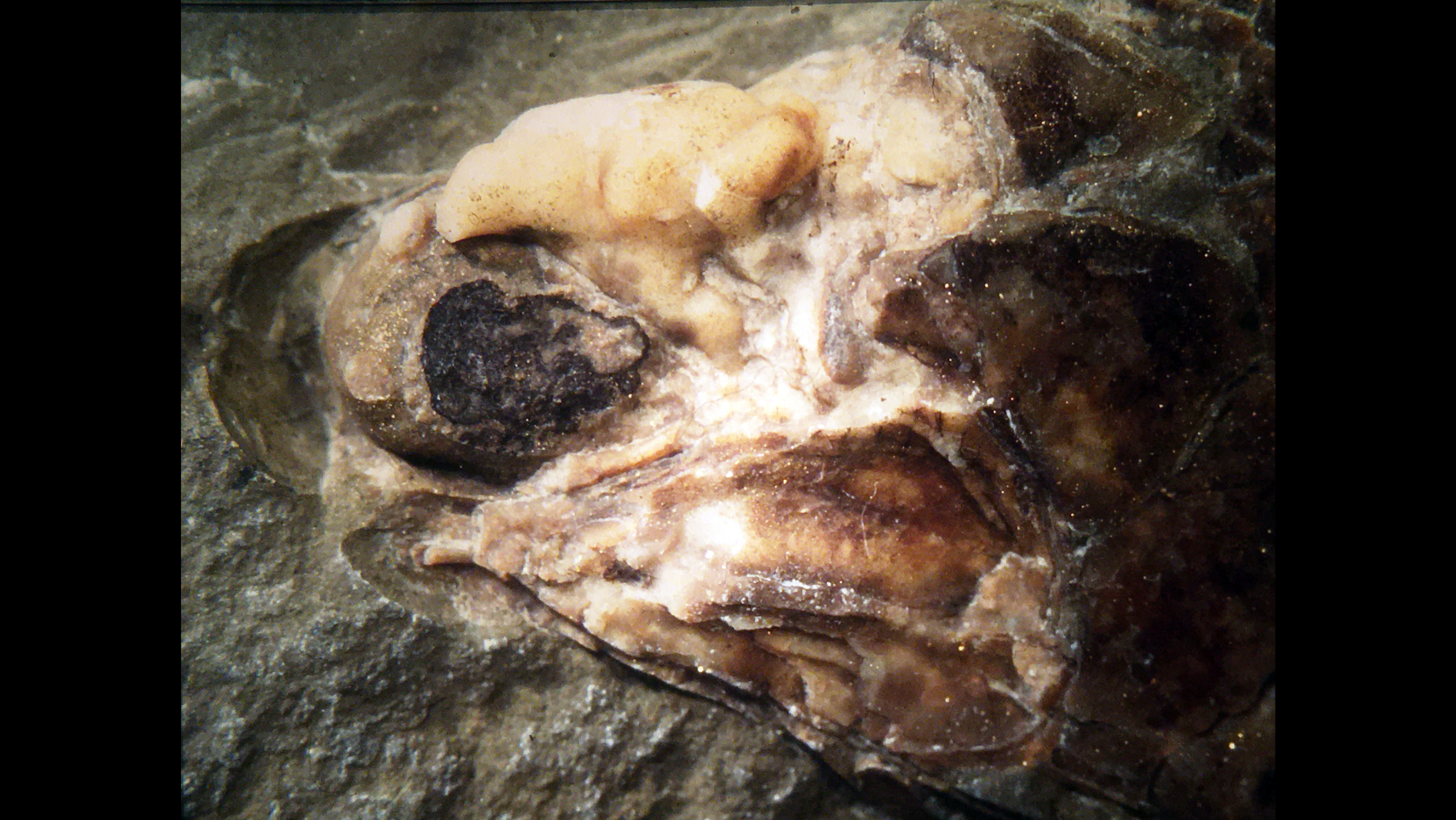
“Soft tissue preservation, in general, is not common in the fossil record, and usually what gets preserved are things like skin or muscles. It's quite rare for neural tissues to be preserved at all because they decay so quickly,” said Abigail Caron, lead author of the study and recent recipient of a doctorate from the University of Chicago, in a statement. “So, the importance of this specimen is that we can now study brain evolution in similar fossils where we only have the bony parts or the infill.”
The fossils' more recent history is also interesting. The Trawdenia specimen was first found in the late 19th century as a round rock nodule that was split in two and ended up at the Natural History Museum in London recorded as two different specimens – until this study's authors realized they were two halves of the same thing.
Using CT scans and 3D models of the skull helped the researchers learn more about the tissues inside the skull compared with the size of the braincase.
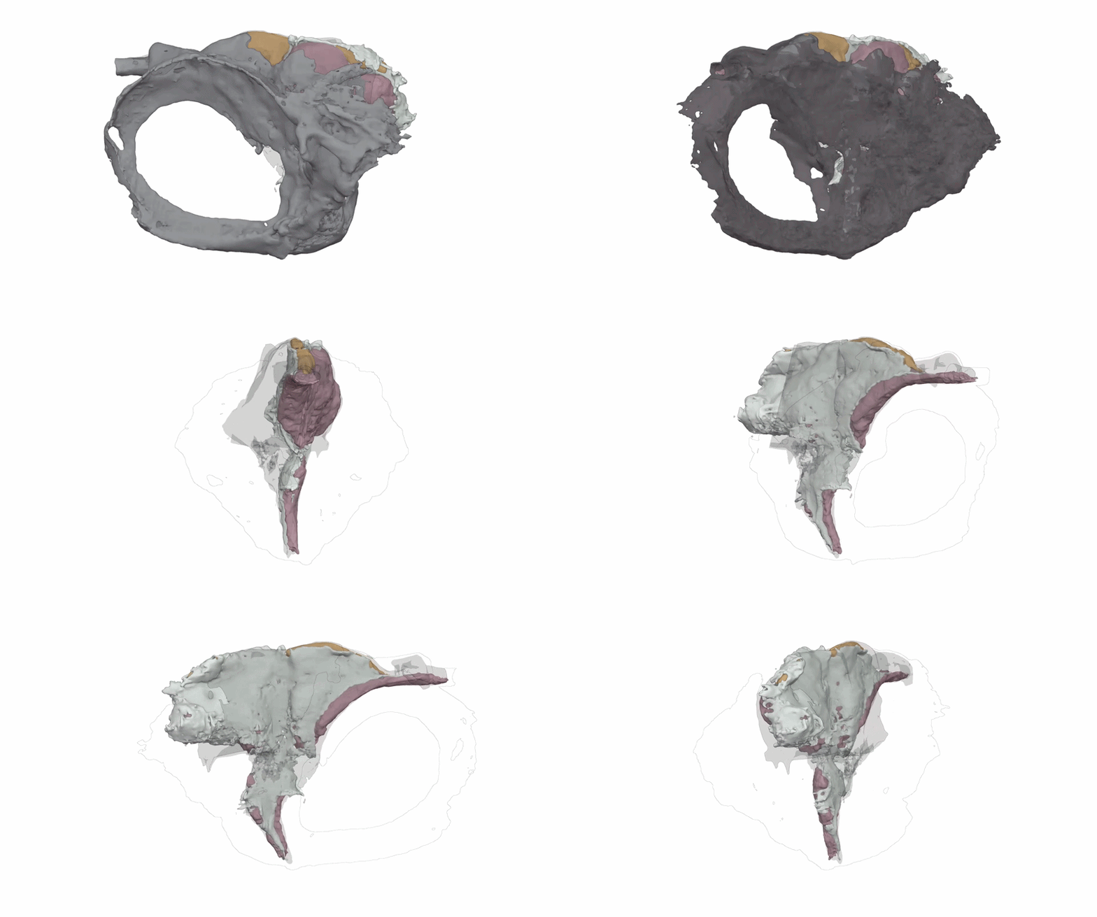
What makes this fossil more surprising is that almost all existing fossil fish brains seem very small in comparison with the amount of space inside the skull, indicating a lot of empty volume. However, in T. planti’s case, detailed scans show that the brain actually sat very snug inside the skull space with very little spare volume.
“What we’re learning by looking at the shapes of the soft tissues is that it's not their relative sizes that matter; it's how they're packed together inside the skull,” said Michael Coates at the University of Chicago, senior author of the new study. “We might be seeing the earliest radiation of fishes that nowadays are represented by paddlefish and sturgeons.”
The paper is published in PNAS.