

Researchers have injected a billion cardiac cells created from stem cells into monkeys in hopes of boosting the regeneration of their injured hearts -- and it seemed to work. This proof-of-principle study means that one day, heart cells beating in a dish would allow survivors of severe episodes to have properly working hearts.
A common type of heart attack called myocardial infarction blocks blood flow in major arteries and deprives muscles of oxygen, weakening the vital pump’s abilities. Infarcted heart muscle doesn’t grow back. Just this week, we learned how genetically modified pigs’ hearts are working fine in baboons. Previous work in rodents have suggested that cardiac muscle cells (cardiomyocytes) derived from stem cells could repair damage. But their hearts beat 400 times a minute -- we’re about 70 beats a minute.
Charles Murry and colleagues from the University of Washington wanted to see if cardiomyocyte transplantation could work in larger primate hearts. They started by producing one billion cardiomyocytes derived from human embryonic stem cells. This was ten times more than researchers have been able to generate. Growing so many cells required a lot of petri dishes, with a total surface area equivalent to 30 medium-sized Domino's pizzas for every monkey, Murry tells Popular Mechanics. "We had incubators stacked floor to ceiling with cell cultures. The actual volume of the cells is about a milliliter and a half, but they need more room when they're growing.”
Then, to create the infarction, they blocked the coronary artery of pigtail macaques for 90 minutes using a catheter and a small balloon. Two weeks later, they delivered the cells into the failing monkey heart muscle. Over subsequent weeks, the cells infiltrated the damaged heart tissue, matured, and assembled into muscle fibers. When they became electrically stable, they began to beat in near synchrony with the primate heart cells, showing “ electromechanical coupling.” After three months, the grafted cells appeared to be fully integrated into the muscle.
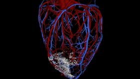
The transplanted cells also remuscularized the failing hearts: New muscle grew up to 0.6 inches wide, and on average, 40 percent of the damaged heart tissue was regenerated. Ultrasound on monkey hearts showed that ejection fraction, which indicates the ability to pump blood, improved in some of the treated animals (though not all). And arteries and veins from the macaque hearts grew into the new heart tissue -- the first time we’ve seen blood vessels from a host animal nurture a large stem-cell derived graft.
In these images, the human cardiac muscle graft (green fluorescent) in the host macaque heart (red) contains myofibril bundles with well formed sarcomeres (left) and a rich network of coronary blood vessels (right).

But, unlike with the rodent studies, some arrhythmias (potentially life-threatening irregular heart beats) were observed in the monkeys -- though they seemed to disappear as the new cells matured electrically. They’ll need to sort this out and also examine the effects of larger areas of infarction before moving into humans. And, since the cells don’t match the patient’s DNA, drugs must be taken to suppress immune system attacks on foreign material.
"The bottom line is, it looks like this works pretty well in a monkey,” Murry says. “And I think if we can do this in a monkey, we can do it in a human." They expect clinical trials to start within four years, and maybe in a decade’s time (with some careful examination of discrepancies), stem cells could be a viable treatment for heart failure.
The work was published in Nature this week.
Images: Murry Laboratory (top), Veronica Muskheli (all others)