

Like many aspects of human biology, there’s plenty about hearing that we’ve yet to understand. Thanks to a new Neuron paper, however, we’re now one step closer to auditory enlightenment.
Sounds travel as a wave. When one is produced, it propagates through a medium that can deform and take its original shape, like air, or water, and reflects off various surfaces, losing energy as it does so. Those are the basics, and fortunately, we’ve evolved, like many other critters, to be able to process it.
Sound leaps into our ears – shaped in such a way that optimizes sound wave capture – and it heads toward our eardrum at the end of the ear canal. This causes the eardrum to vibrate, which ends up shaking a very delicate, tiny collection of bones. These bones then shake a fluid-containing segment named the cochlea, which initiates a response in the auditory nerve.
The signal, now electrical, gets sent to our brain, where it’s quickly processed and we understand it as noise. Those are the basics, but there’s plenty of blank spaces that scientists have yet to fill in.
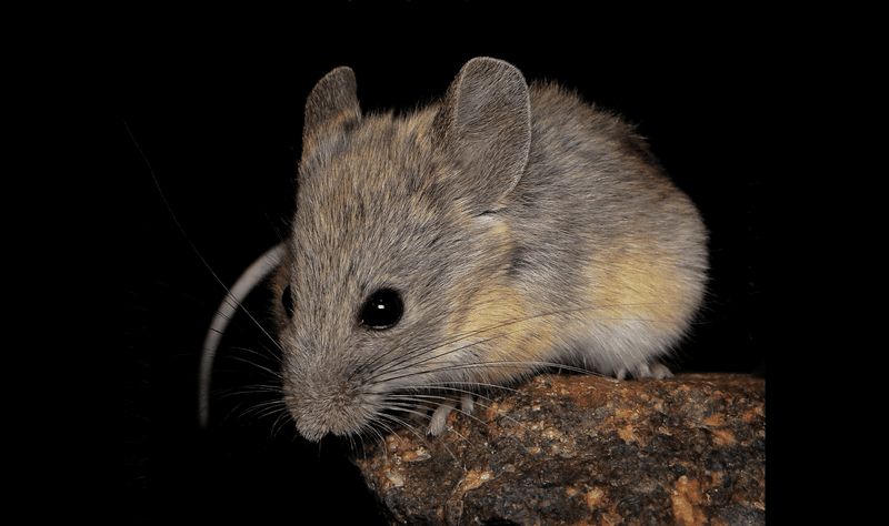
Take the extremely tiny hairs in your ear, for example. We’re not talking about those in the ear canal, by the way – they’re designed, along with earwax, to keep dirt, debris, and hostile microbes away from your middle and inner ears.
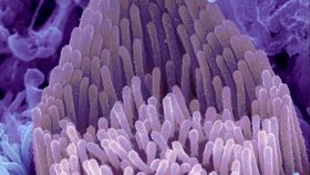
You also have incredibly fine slithery things inside your inner ear too, within the snail-shell-shaped cochlea. Within a segment known as the Organ of Corti, you have hair cells, adorned with hair-like structures named stereocilia.
When vibrations reach them, these structures also vibrate, and it’s these movements that are converted to nerve impulses for the brain to comprehend. This is a type of process known as mechanotransduction, which describes how cells receive physical forces and turn them into biochemical and biological responses.
As spotted by The Scientist, this new paper, led by Boston Children’s Hospital and Harvard Medical School, answers a key question about those stereocilia. Namely, what proteins drive the conversion of their mechanical, vibrational movement into electrical signals?
Researchers have been trying to answer this question for decades now, but the problem is that identifying this protein requires either replicating the complex biological hearing apparatus in a laboratory setting, or observing the protein do its thing in a living person, or animal.
Both have proved to be tricky, to say the least. In order to narrow things down, the team conducted an incredibly delicate series of experiments.
They created multiple mutated variants of TMC1 proteins, the prime suspects as it were, and injected genes responsible for them into mouse younglings that lacked them. After allowing the hearing organs to begin to take shape, they then extracted them, and let them grow in a lab setting for a little longer.
The team were looking for evidence that TMC1 acted as a bridge, one that permitted electrically-charged atoms, or ions. Such a bridge, known as an ion channel, would permit the full conversion of stereocilia movements into nerve signals.
By taking careful measurements, and using various compounds to interrupt their electrochemical properties, the team confirmed that TMC1 was in fact the ion channel they were looking for. Our understanding of hearing isn’t exactly complete, and it’s likely multiple proteins are involved, so the story’s not over yet. Nevertheless, this paper represents a major advance in the field. Progress is – ahem – earreverseable at this point.
[H/T: The Scientist]