

Using human stem cells, researchers have created three-dimensional, light-sensing eye tissue that can be used as a study system for understanding diseases that cause blindness.
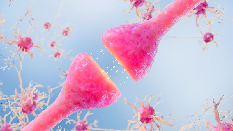
The retina is the light-sensitive layer of tissue at the back of our inner eye. It’s like the film in a camera: Images come in through the lens and focus on the retina, which then converts the images into electric signals to the brain. Many forms of blindness result from the dysfunction or loss of the retina’s light-sensing cells, called photoreceptors. Researchers have created retina-like structures in the past, though these such structures haven’t been shown to sense light or transmit impulses to the brain.
Now, researchers have created retinal tissue that resembles the anatomy of the developing eye, containing photoreceptors that respond to light the way ours do in real life. “We have basically created a miniature human retina in a dish that not only has the architectural organization of the retina but also has the ability to sense light,” M. Valeria Canto-Soler from Johns Hopkins University School of Medicine says in a press release.
Canto-Soler and colleagues developed the light-sensing retinal tissue from human induced pluripotent stem (iPS) cells. These are adult cells that have been genetically reprogrammed to their primitive state. Under the right conditions, iPS cells should be able to develop into any of the 200 or so cell types in the human body. Here, the team turned them into precursor cells destined to become retinal tissue. The growth of the cells, and the subsequent tissue, in the petri dish corresponded in timing and duration to retinal development in a human fetus.
“We knew that a 3-D cellular structure was necessary if we wanted to reproduce functional characteristics of the retina,” Canto-Soler says, “but when we began this work, we didn’t think stem cells would be able to build up a retina almost on their own. In our system, somehow the cells knew what to do.”
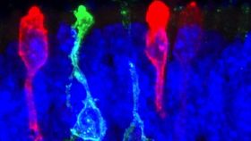
When the mini-retinas reached a stage equivalent to 28 weeks in the womb, the team tested the photoreceptors by placing an electrode into a single cell. When they sent a pulse of light to the cells, they reacted in a biochemical pattern similar to photoreceptors in people who are exposed to light. In particular, they acted like our rods, which enable vision in low light. (Cones, the other type of photoreceptor cell, respond to bright light.) Pictured here: Photoreceptors derived from human iPS cells in a dish can detect light.

But this new retinal tissue isn’t meant to be used as transplants, at least not yet. For one thing, it doesn’t produce visual signals that the brain can interpret into an image. The lab retina is more of a complement of human retinal tissue that can help model diseases -- such as retinitis pigmentosa -- to help develop technologies for restoring vision.
The work was published in Nature Communications this week.
[Johns Hopkins Medicine via The Scientist]
Images: X. Zhong. C. Gutierrez and M.V. Canto-Soler at the Wilmer Eye Institute, Johns Hopkins University School of Medicine