

It’s a beautiful thing when science and art brush aside their superficial differences and join together to celebrate the magnificence of this amazing world.
The rest of this article is behind a paywall. Please sign in or subscribe to access the full content.Nikon’s Small World Photomicrography Competition is a great example of this. The competition has been running since 1975 to recognize and applaud the innovations in photography of teeny weeny things, using microscopes. This year had more than 2,000 entries from 83 different countries and the images were as spectacular as ever.
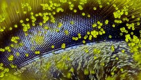
This year’s winner was Ralph Grimm, a self-taught photomicrographer from Australia who works as a school teacher. His winning piece was a 120x magnified photo of a honey bee’s eye covered in single grains of dandelion pollen. The image took over four hours to set up, with Grimm having to painstakingly mount the eye, carefully set the focus and illuminate the subject without any smudging.
The image also has a social message. Grimm was formerly a keen beekeeper and wanted to use his skills to highlight the plight of the world’s bees. On winning the award, he said: “It’s a subject of great sculptural beauty, but also a warning – that we should stay connected to our planet, listen to the little creatures like bees, and find a way to protect the earth that we all call home.”

Image credit: Kristen Earle, Gabriel Billings, KC Huang & Justin Sonnenburg/Nikon
Kristen Earle, Gabriel Billings, KC Huang and Justin Sonnenburg came close to winning with their microscopic photo of a mouse’s colon (above). They are a team of microbiologists from the Stanford University of Medicine. The image shows a thriving community of bacteria (shown in red) being separated from the colon tissue (blue) by a layer of mucus (green).

Image credit: Henri Koskinen/Nikon
Henri Koskinen from Helsinki, Finland, took a photo of a spore capsule of some moss. It may not sound too miraculous, but the photo shows a ridiculous amount of detail of this microscopic entity.

Image credit: Richard Kirby/Nikon
Richard Kirby snapped a 450x magnified picture of the planktonic larva of a horseshoe worm at the Marine Biological Association in Plymouth, England.

Image credit: Evan Darling/Nikon
Taken with a 10x magnification, Evan Darling took a photograph of a microscopic starfish at Memorial Sloan Kettering Cancer Center in New York.

Image credit: Ian Gardiner/Nikon
Ian Gardiner's image is even more miraculous when you realize it is of a live creature. Taken with a 25x magnification, this photo shows a clam shrimp (Cyzicus mexicanus).
These are just a select few of the entries, all of which you can view on the Nikon Small World website. Nikon also showcases its winning entries in a tour of museums in North America. So far, exhibitions have been announced at Tellus Science Museum In Georgia, Indiana State Museum, Brooklyn Children’s Museum, New York State’s Rochester Institute of Technology, Adventure Science Center in Tennessee and Science World in Vancouver.