
Ad

health-iconHealth and Medicine
clock-iconPUBLISHEDMay 25, 2016

It May Look Pretty, But The Zika Virus Is Far From Pleasant

Tom Hale headshot

Tom Hale

Tom has a Master's degree in Journalism. His editorial work covers anything from archaeology and the environment to technology and culture.

Senior Journalist

Tom has a Master's degree in Journalism. His editorial work covers anything from archaeology and the environment to technology and culture.View full profile

Tom has a Master's degree in Journalism. His editorial work covers anything from archaeology and the environment to technology and culture.

View full profile
article image
David S. Goodsell/RCSB Protein Data Bank

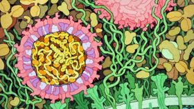
In a beautiful illustration, David Goodsell from the Protein Data Bank renders the structure of the Zika virus visible in order to show how this virus wreaks havoc on the human body and its cells.

The rest of this article is behind a paywall. Please sign in or subscribe to access the full content.

Scientists discovered the Zika virus in 1947, but they have only recently revealed its near-atomic structure via cryo-electron microscopy. The structures in the artistic rendering show that Zika is very much like other flaviviruses, the genus of which includes West Nile, dengue, and yellow fever.

The Zika virus itself can be seen in the two pink circles, one of which is shown in a cross section. In its core, you can see the RNA genome (yellow) with capsid proteins (orange). Its outsides show envelope proteins (red) and membrane proteins (magenta). Most interestingly, it shows the virus interacting with receptors on the cell (green), and surrounded by blood plasma molecules at the top.

Image credit: David S. Goodsell/RCSB Protein Data Bank

[H/T: Gizmodo]


Written by 

Add us as a Google preferred source to see more of our
trusted coverage in Search