

A new implantable tissue scaffold plastered with multiple growth factors can help regenerate bone tissue that looks and behaves like the original. So far, it has helped repair traumatic skull injuries in rats. The work was published in Proceedings of the National Academy of Sciences this week.
The current standard for treating bone injuries is to transplant bone from another part of the patient’s body. This painful process doesn’t always supply enough bone for the severe injuries suffered by soldiers or patients with congenital defects. “It’s been a truly challenging medical problem,” says Nisarg Shah of MIT.
For natural wound healing, two of the most important bone growth factors are the platelet-derived growth factor (PDGF) and bone morphogenetic protein 2 (BMP-2). After a fracture, the body releases PDGF immediately, followed by other factors like BMP-2, which recruit stem cells to the injury site in order to produce bone and form support structures, such as blood vessels.
The amount and timing of growth factors is key: Large quantities delivered too quickly will be rapidly cleared away from the treatment site. So Shah and colleagues looked for a way to release small quantities (nanograms, not milligrams) of growth factors slowly over several days or weeks.
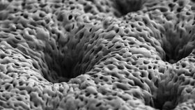
They started by coating a thin, porous sheet of polymer with layers of growth factors: 40 layers of BMP-2, topped with 40 layers of PDGF. Just like the body’s natural healing process, this polymer multilayer allows for PDGF to be released quickly in just a few days, along with a more sustained BMP-2 release. The polymer membrane (called PLGA, pictured below) can be programmed to disintegrate at certain rates, so it stays in the body for only as long as it's needed.

The team implanted the scaffold in rats with a large skull defect about 8 millimeters wide -- something that definitely couldn't heal on its own. The two growth factors worked together to mobilize precursor cells to the wound to form new tissue. Within two weeks, a layer of bone was generated, and the researchers say it’s indistinguishable from natural bone in both its appearance and mechanical properties.
“Using this combination allows us to not only have accelerated proliferation first, but also facilitates laying down some vascular tissue, which provides a route for both the stem cells and the precursor osteoblasts and other players to get in and do their jobs,” MIT’s Paula Hammond says in a news release. “You end up with a very uniform healed system."