


 Health and Medicine
 PUBLISHED


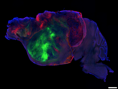


Scientists often want to study how the human brain develops and how different conditions can impact its journey, but cracking open a skull and having a peek isn’t always a viable option. To get around this problem, a team from Stanford Medicine has developed a new way to transplant human mini-brain organoids into the skulls of mice.
A new brain: part mouse, part human
The researchers used bioengineered mice that are essentially born without most of their cortex, the outermost layer of the brain that handles high-level thinking, memory, language, and decision-making.
They created these edited rodents by tweaking their genes to eliminate the cells that would normally form those brain regions during early development.
Although the cortex-depleted mice performed slightly worse in tasks that tested their memory and fine motor skills, they were otherwise quite typical – which itself is remarkable.
“These animals – that lack half of the volume of the brain or 98 percent of the cortex and hippocampus – are surprisingly functional,” Sergiu Pașca, study author and professor of psychiatry and behavioral sciences at Stanford University, told reporters at a media conference this week.
“You wouldn't be able to tell which is a [cortex-depleted] mouse from the others if you were just to look at them, which really tells us that if you remove parts of the nervous system very early on in development, there is remarkable plasticity of the rest of the nervous system that can presumably take over,” Pașca added.
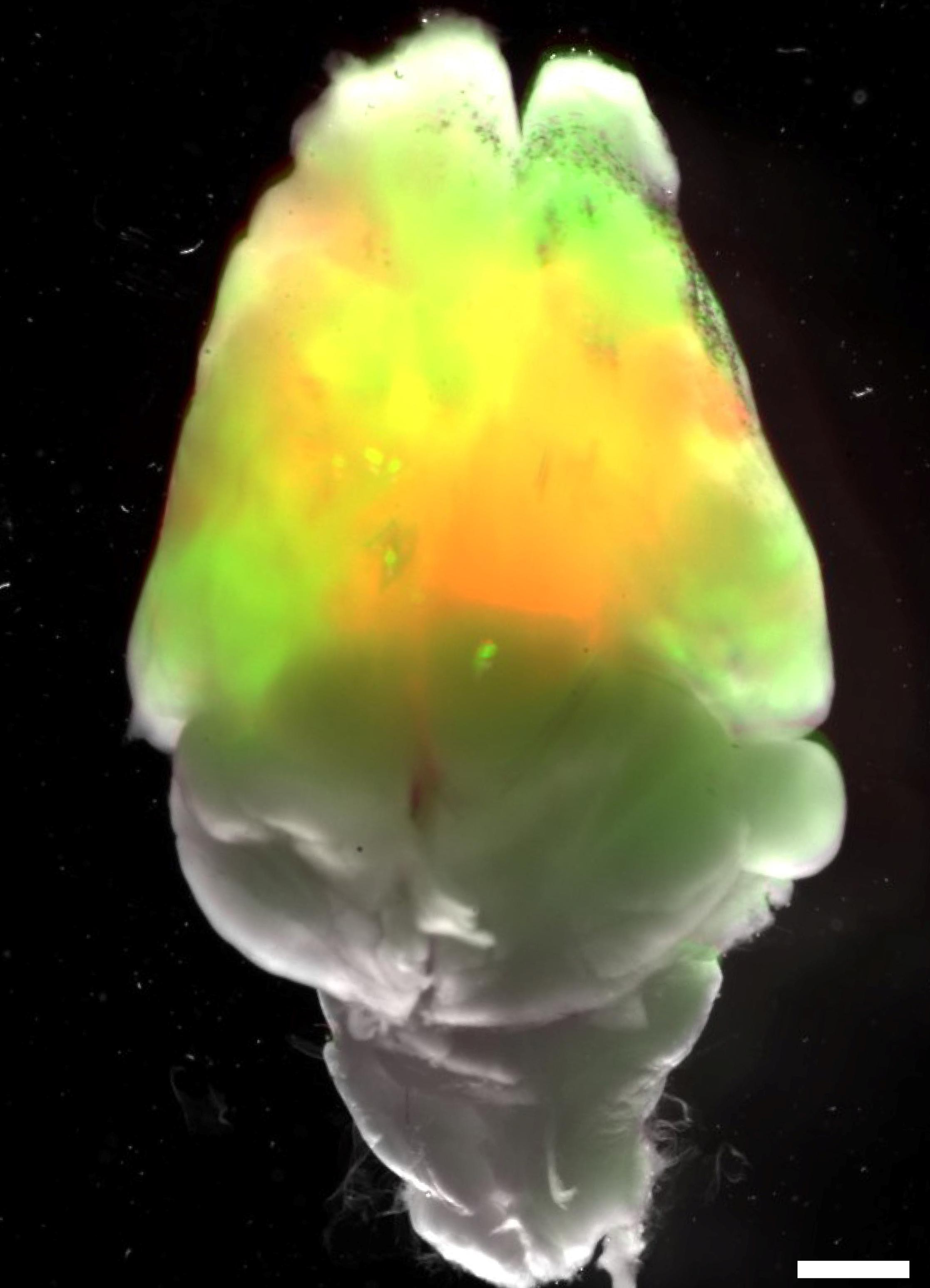
Utilizing the empty space in the skull, they surgically transplanted a three-dimensional bundle of neurons grown from human cells.
Scientists use these miniature human brain organoids as a proxy to study the real deal, but this new feat allows scientists to see how they behave in a living, breathing organism, not just a glass Petri dish.
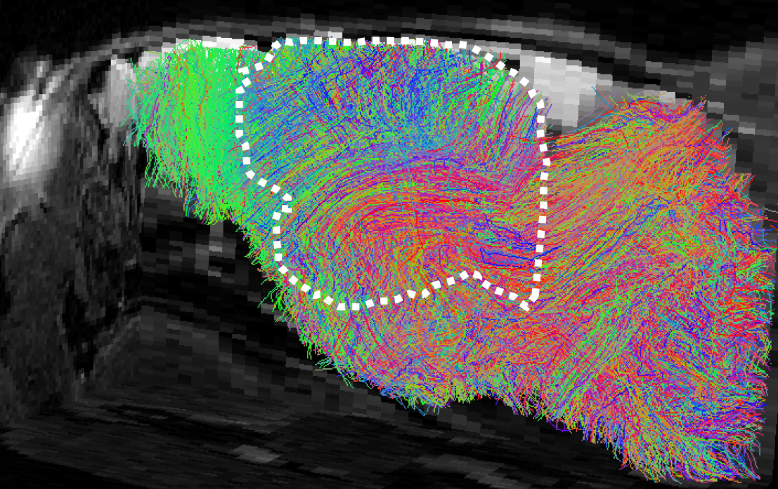
Remarkably, the human brain tissue thrived inside the “xenocortical mice”, expanding in volume almost five-fold within three months. The human organoid also developed working connections with the native brain and spinal cord.
“These human cells are relatively well connected because they have more space and more time with the rest of the nervous system. They're electrically active. You can actually probe them and see how the cells are forming some of these networks,” explained Pașca.
How the xenocortical mice could help to understand human brains
The next part of the study explored whether xenocortical mice could serve as a model for studying how various diseases and developmental conditions affect the human brain.
For instance, the human brain is extremely susceptible to low oxygen during development, much more so than the mouse brain. This vulnerability is a major contributor to conditions like cerebral palsy.
After five hours of low-oxygen exposure, the xenocortical mice showed significant damage to their human-derived cortical tissue, just as one would expect. These oxygen-deprived mice also struggled with moving and maintaining balance, echoing the symptoms of cerebral palsy in humans.
Meanwhile, the “normal” mice were almost totally unfazed by the low-oxygen experience.
“Finding out what accounts for this difference could yield clues about human neural susceptibility to oxygen deprivation, shed light on mechanisms underlying cerebral palsy and provide a platform for testing potential therapeutic strategies,” Pașca said in a statement.
The researchers go on to explain that the xenocortical mouse models could be used to study the origins of other human conditions or neurodevelopmental disorders, including schizophrenia, epilepsy, autism, and more.
Most incredibly, the human mini-brain that’s transplanted into the mice could be derived from a specific person’s DNA, allowing the researchers to essentially track how and why an individual's brain functions the way it does.
“The cells we implant carry the genetic material of the person they’re derived from — whether that person is a patient or a healthy individual — allowing us to study downstream disease effects in brain cells and circuits,” Pașca said.
A bold breakthrough, with some limitations
Other researchers in the field have praised the study, suggesting it's a promising new way to probe the mysteries of human brain development.
However, they've also cautioned that the model has real limitations. Chief among them, it will be tricky to disentangle normal human brain development from the effects of growing up in the unusual environment of a xenocortical mouse brain.
“[W]e should be cautious here: the functional integration of human-derived brain cells is not a proxy for its functional equivalence. The grafted human brain tissue lacks features of mature human brain tissue. And the recorded behaviour emerges from the interactions of the human cortex and the remaining mouse subcortex,” Adeel Razi, Professor of Computational Neuroscience at Monash University, who was not involved in the study, said in a statement to the Australian Science Media Centre.
“Hence the limitation of the technique is that it is difficult to know whether the grafted human brain cells are necessary or sufficient for a particular observed behaviour,” commented Razi.
The study is published in the journal Nature.



 1
1