

When humans suffer from liver failure, the organ’s capacity to regenerate itself is limited by surrounding scar tissue that stresses the cells and causes them to die. A new study published in the journal Liver Transplantation has discovered a potential solution, as they found that pig’s liver cells were able to grow in their lymphatic system. If a significant enough volume of cells can establish themselves ectopically in this way, it’s possible this unusual approach could contribute towards better treatments for liver failure in humans.
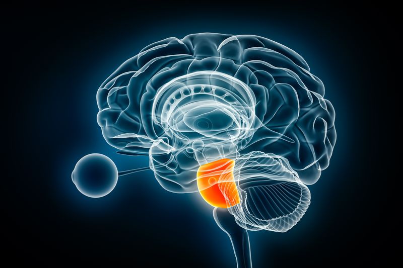
The liver contains hepatocytes, which are the chief functional cells of the organ and are capable of regenerating. They do this normally but need a healthy environment in which to do it, but in the late stages of liver failure the organ becomes too scarred and toxic for the hepatocytes to thrive. Instead, the cells die and without a liver transplant so too will the patient.
In his previous research, Eric Lagasse, an associate professor of pathology at the University of Pittsburgh School of Medicine, noticed that liver cells could regenerate if they were injected into the lymph nodes of mice. When enough hepatocytes were established, the auxiliary organ could compensate for mice with a genetically-induced malfunctioning liver. The discovery was promising but the livers of mice are small and Lagasse wasn’t sure if the same result could be replicated in a larger animal.

Their most recent study set out to trial the therapy on pigs whose livers had been surgically altered to mimic human liver disease. They extracted hepatocytes from a healthy sample of the pig’s liver and injected these into the abdominal lymph nodes of the same pig they were taken from.
Of the six pigs operated on, all showed signs of recovery from liver failure. Inspection of the ectopic hepatocytes revealed that not only were they thriving, but they had established a network of bile ducts and blood vessels and grew bigger as the pig’s original liver lost function. This compensatory growth seems to indicate the pig’s bodies were maintaining an equilibrium of liver function as an alternative to continuously churning out more and more hepatocytes in a growth rate comparable to cancer.
Lagasse believes that if successful in humans, the auxiliary livers could compensate for liver failure regardless of the cause, from hepatitis to alcoholism, and will soon begin a human clinical trial for the treatment.