

Eighty years after the last Tasmanian tiger (Thuylacinus cynocephalus or thylacine) died, we are still learning about this species from museum specimens collected at the time. Using a novel method to reconstruct the white matter lost from preserved brains, two scientists have discovered a lot about the construction of the thylacine's brain, suggesting the extinct creature had a well-developed planning capacity.
Thylacines once roamed across much of Australia and New Guinea. By the time Europeans arrived in Australia however, they were restricted to Tasmania, and were easily the largest surviving marsupial carnivore. A bounty for their corpses and competition with wild dogs saw them die out by 1936, although rumors of wild survivors still persist.
Early Australian scientists looked down on thylacines, as they did on other Australian native species, regarding marsupials as inferior to placental mammals. However, Emory University's Professor Gregory Berns and Professor Ken Ashwell of the University of New South Wales have challenged that notion, showing the thylacine's brain was well developed in regions responsible for planning and decision making.
Berns and Ashwell made MRI scans of two thylacine brains preserved more than 100 years ago and held at the Smithsonian and Australian museums, and used diffusion-tensor imaging (DTI) to fill in the parts made from material more prone to decay. They compared these with scans of preserved brains of the thylacine's closest living relative, the Tasmanian devil. The results have been published in PLOS ONE.
Allowing for their greater body mass, thylacine's brains were 25 percent larger than a devil's. This was particularly notable in the caudate zones, which the authors consider consistent with the suspicion they were ambush predators, rather than scavengers, with the capacity for strategy.

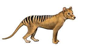
Compared to its nearest living relative, the thylacine's brain was larger, more compartmentalized, and more developed in areas responsible for planning. Berns and Ashwell/ PLOS ONE
Ashwell noted in a statement that the study is as significant for its methodology as for the findings. “MRI imaging of the preserved brains of rare, extinct and endangered species is an exciting innovation in the study of brain evolution. It will allow us to track pathways and study functional connections that could never be analyzed using older experimental techniques.” Previous efforts have used much fresher specimens of surviving species.
Despite very different evolutionary histories, thylacines' skeletons came to resemble dogs so closely they were used to trick zoology students in Oxford University anatomy exams. Their brains, however, were structured very differently.
“The natural behavior of the thylacine was never scientifically documented, and what we know comes from observation of zoo animals, whose behavior is not indicative of wild animals, and stories from trappers,” Berns said.
Once deliberately driven to extinction, thylacines have become symbolic of the island they inhabited and the natural world that is disappearing. The preservation efforts of museums have at least ensured we didn't lose all knowledge as well.