

The complexity of bumblebees' behaviour is at odds with their puny brains. While their brains are just 0.0002 percent the size of a human's, they’re capable of some surprisingly high levels of communication, memory and navigation. By using newly developed computed tomography (CT) scanning techniques, scientists from Imperial College London might have begun to lift the bonnet on bees' cognitive abilities.
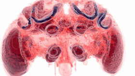
The rest of this article is behind a paywall. Please sign in or subscribe to access the full content.The researchers used micro-CT imaging to detail the brain structure of 19 bumblebees (Bombus terrestris) in a study recently published online in Scientific Reports.
They used the micro-CT scanner facility at the Natural History Museum in London. This technique uses X-rays and computer technology to capture hundreds of “image slices” of the subject, which can then be compiled to create an accurate 3D projection. Unlike the old techniques of analyzing insect brains with scalpels and decapitations, it’s non-invasive and a lot less fiddly.

CT scan of bee brain tissue. Dylan Smith/Imperial College London
“It’s a fantastic way to look inside insect brains. We can look at the brain as it naturally sits in the bee’s head, without the human error of having to extract it,” Dr. Richard Gill, one of the researchers from the Department of Life Sciences at Imperial, said in a statement.
“The 3D structures can also be explored as you wish – from looking at the whole organ down to each separate structure, piece by piece.”
Aside from creating some beautiful images, understanding the mechanics of bees' brains has some far-reaching potentials. Bees play an essential role in ecosystems across the world as pollinators. Despite this, changes to habitats and environments has seen a worrying decline in bee populations over recent years. By practising this more precise art of CT scanning, they hope to better understand outside influences on bees' brains – such as trauma and disease – and how this affects their ability to navigate and pollinate.
“The structures are so small that tiny errors in measurement can lead to wrong conclusions. This new technique allows structures to be isolated, examined, and measured in greater detail than ever before,” said Dylan Smith, another author of the paper.

Scanning allows for greater detail and precision in isolating parts of the brain. Dylan Smith/Imperial College London

The external head case showing brain in situ. Dylan Smith/Imperial College London
[H/T Motherboard]