

A team from The Rockefeller University in New York have transplanted human stem cells onto newly formed chicken embryos in the lab, forming dual-species clusters called chimaeras that were observed for the next 24 to 48 hours of growth.
The experimental method, described in Nature, has already provided key insight into early developmental processes that had been previously impossible to study. And after demonstrating a high degree of consistency and reproducibility, the authors suggest that their technique holds promise to facilitate even more pioneering research.
Published online yesterday, their paper has, of course, already been vilified in the press. But what did they really do, and why?
Soon after fertilization, vertebrate embryos transition from a simple hollow clump of cells to a three-layered clump of cells that has a defined head-to-tail, back-to-front, and left-to-right axis. To the untrained eye, the embryo still looks like a vague aggregation of cells following this step, far removed from the organism it is going to become; yet in reality, the development plan for the entire body’s layout and all its diverse tissue types has been set in motion.
Decades of animal investigations have revealed that the driving force for this step is specialized “organizer cells” that function similarly across species.
But thanks to a strict ethical ban on culturing human embryos past post-fertilization day 14, developmental biologists have been unable to watch how organizer cells arise in humans – thought to occur soon after this point – and mediate the crucial three-layer phase that, in a natural pregnancy, would unfold immediately after implantation. Such knowledge is also important for reproductive science, as it is during this period of cell layer migration and rearrangement that most miscarriages occur.
“No one knew what happens after the ball of cells attaches itself to the uterus,” lead author Ali Brivanlou told Nature.
Hoping to take the first step toward recreating a human organizer, the Rockefeller team grew colonies of human stem cells using certain growth factors and a forced culture shape, causing them to adopt the features of an early embryo. These clusters were then transplanted onto chicken embryos that were removed from eggs at the 12-hour mark, a point that corresponds to day 14 in a slower-growing human embryo.
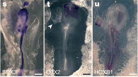
The stem cell colonies were then transplanted onto the chicken embryos at a site that later develops into neural tissue. Remarkably, in the brief time window after the hybrids were created, the human cells induced the formation of a secondary axis in the chicken cells and began to direct them to develop a second chicken nervous system – thus confirming that the replication method was successful.
(Note: The team limited human cells to less than 10 percent of all cells in the chimaera embryos and none were allowed to develop past 48 hours.)
“It’s a real advance – it’s beautiful this can be shown without the need of using embryos,” commented German developmental biologist Martin Blum. “At the moment I could not think of a case where an early human embryo would be needed to answer basic questions.”
Brivanlou himself disagrees, stating there is no replacement for studying actual human embryos.
“Human embryonic stem cells and eggs have all the information,” he said. “Everything else we do when we try to model kind of oversimplifies it.”
Nevertheless, a torrent of follow-up research is sure to come.