

Imagine if a trip to the dentist to treat a cavity didn’t involve a filling, root canal, or crown. What if a simple light treatment could actually get your teeth to regrow themselves using stem cells? That’s the aim of a group of researchers at Harvard’s Wyss Institute, led by David Mooney, who have found success in regrowing rat teeth in this manner. The researchers have developed a technique using a low-power laser to coax stem cells into reforming dentin, which could have implications for dentistry, wound healing, and bone restoration. The results of the study have been published in the journal Science Translational Medicine.
Proteins known as growth factors are what cause stem cells to differentiate into whatever type of cell they are bound to become. Introducing different growth factors force the cells to develop the desired type of tissue. Unfortunately, it isn’t quite as simple as it sounds. Most of the developments in using stem cells in regenerative medicine have regrown tissues in vitro and later need to be transplanted into the person. This involves a lot of technical care and is a highly regulated process, which slows down progress. Mooney’s team claims they have come up with a new technique that could streamline the process, making it a viable clinical option much more quickly.
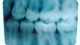
The team set up a miniature dentist office-like setting for the rodents used in the study. They drilled holes into the rats’ molars to simulate tooth decay. Next, adult stem cells were applied to the pulp of the tooth and a non-ionizing, low-level laser was used to stimulate the growth factors. The teeth were then sealed with a temporary cap to be worn over the next 12 weeks. The follow-up x-rays and microscopy analysis showed that the dentin, the layer under the visible enamel, had indeed begun to grow back due to the laser/stem cell therapy.
“Our treatment modality does not introduce anything new to the body, and lasers are routinely used in medicine and dentistry, so the barriers to clinical translation are low,” Mooney said in a press release. “It would be a substantial advance in the field if we can regenerate teeth rather than replace them.”

The researchers used high-resolution x-ray imaging and microscopy techniques to assess the formation of reparative (tertiary) dentin 12 weeks after the low power laser treatment. In the microscopy images shown here, the yellow hashtags (#) sit atop the newly-formed tertiary dentin; there is more tertiary dentin in the laser-treated teeth than in the control. [Credit: Harvard’s Wyss Institute and SEAS]
Of course, performing dentistry on rats was not without its challenges. While the dentin was incredibly similar to that which grows naturally, it wasn’t organized exactly the same way. Also, restored dentin forms what is known as a “dentin bridge” that covers the exposed dental pulp. While this is somewhat easy to detect in human teeth, it was very difficult to see in the tiny rat teeth. Mooney stated that “[t]his is one of those rare cases where it would be easier to do this work on a human.”
The team then sought to identify which molecular mechanisms were influenced by the laser. Transforming growth factor beta-1 (TGF-β1), a widely multifunctional protein that regulates cell proliferation and differentiation, was largely responsible for regrowing the dentin. The laser first stimulated reactive oxygen species (ROS), which has an important function in cell signaling and other cellular homeostatic processes. ROS then stimulated the then-dormant TGF-β1 into activating, which gave the stem cells the signal to differentiate into dentin. The researchers also noted that the reaction was dose-specific to the level of light received.
Anecdotal evidence about the power of low-level light therapy has been piling up for nearly 50 years, but this study was the first to nail down the molecular mechanism. This could open up a host of potential avenues of treatments that expand far beyond dentin. The team’s future research will include experimentation with other stem cells, and they also hope to begin human trials for restorative dentistry soon.
[Header image: “I put the X in X-ray” by Bill Selak via flickr, used in accordance with CC BY-ND 2.0]