

Researchers at the University of California Berkeley have constructed the first detailed map of the 3D structure of telomerase – a hotly studied DNA-safeguarding enzyme thought to play a role in cancer and longevity – following decades of frustrating attempts.
The breakthrough, published in Nature, will open the door for a deeper understanding of cell mortality and could lead to the development of a new generation of treatments for aging-related diseases and cancers.
“It has been a long time coming. It took a lot of persistence,” study lead Kathleen Collins, a professor of molecular and cell biology who has worked on the enzyme for 26 years, said in a statement.
First discovered in 1985, telomerase protects the chromosomes within a cell from becoming damaged following the process of replication that occurs prior to division.
Every time the DNA is copied, short sections of nucleotides at the end of each strand are lost because the enzymes that string together new copies of the DNA template cannot reach all the way down. To protect important gene-encoding sequences from suffering this fate, chromosomes in complex organisms are tipped with repeated sequences of filler nucleotides, collectively referred to as telomeres. Telomerase maintains telomeres by re-adding the repeats after replication.
Yet after embryonic development is complete, this protein is only expressed in certain cells of the body such as germ cells, skin cells, and adult stem cells. Most of the cells in an adult human don’t produce telomerase, and thus, the telomeres within get shorter and shorter with each wave of cell division.
Eventually, the telomeres reach a critical length wherein the integrity of important DNA sequences is at risk. At this point, called senescence, the cell stops dividing in order to prevent faulty copies – which could become cancerous – from being made.
The increase in senescence that occurs as we get older has been associated with signs of aging, such as gray hair, reduced organ function, and impaired wound healing, though it is not known whether short telomeres cause the physical decline of aging to begin or if they are a symptom of it themselves.
And as with any biological process, to understand telomeres we first need to mess with them. Such experiments require compounds that can mimic or alter the action of telomerase, and these can't be created without precise structural knowledge.
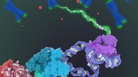
But until now, imaging studies have been unable to achieve the level of resolution necessary to tease out the structure and interactions of the many protein subunits that move about on the molecule’s RNA backbone.
So, the team turned to a new, Nobel Prize-winning form of microscopy, called Cryo-EM, that fires beams of electrons at organic molecules in a frozen state. Cryo-EM is ideally suited for complex, multipart proteins such as telomerase.
“The best previous images of human telomerase had a resolution of only 30 Ångstroms [ten billionths of a meter]; we were able to get about 7 to 8 Ångstroms resolution using cryoelectron microscopy,” Kelly said. “When I got to the point where I could see all the subunits – we had 11 protein subunits in total – it was a moment of, ‘Wow, wow, this is how they all fit together.’”
According to the team’s press release, they have already begun modifying their technique in order to get the resolution of 3-4 Ångstroms needed for drug development.