

Advancements made in a revolutionary electron microscope technology has enabled researchers to break the atomic resolution barrier and image individual atoms within a protein. This is a massive step forward for the process, called cryo-electron microscopy, which has never been able to produce images at such high resolution before.
Cryo-electron microscopy has been rapidly revolutionizing structural biology ever since three scientists won the Nobel Prize in Chemistry for developing it to a high resolution back in 2017. Since then, improvements in electron beam technology alongside software optimizations have allowed it to go deeper into molecular structures than ever before. Peering into the atomic structure of proteins will push forward our understanding of cellular processes immensely, whether that be enzyme interactions or visualizing drug binding.
The technology is now nipping at the heels of the more widely used X-ray crystallography, a complex process that can sometimes take months to complete or, for some proteins, may not produce an image at all.
To image proteins at such detail, cryo-electron microscopy requires some pretty cold temperatures. After taking proteins and cooling them to cryogenic temperatures (around -200°C), an electron beam is applied to the sample. Normal electron microscopy requires complex sample preparation, but with cryo-electron microscopy, all that is needed is the sample to be frozen solid. Electrons barrage the sample and bounce off back into a detector, which then sends the data through computer software to make sense of it. The software stitches together the final image, and you can go about studying the structure of your protein.
Reporting in Nature, a team has now managed to image a protein at such a high resolution that all the atoms in it were clearly visible. This is the first time individual atoms in a protein structure can be clearly made out in a cryo-electron microscope image.

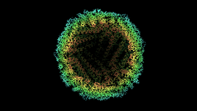
The protein, called apoferritin (usually found as ferritin, an iron-storing molecule), the image captured had an astonishing resolution of 1.25 ångströms – smashing the previous records of 1.54 ångströms. Apoferritin is used in cryo-electron microscopy due to its extreme stability and has become a reliable test sample for pushing the technology to its limits.

As breakthroughs into the technology continue, the possibilities are endless as to how cryo-electron microscopy will shape structural biology. However, it is looking like we are reaching the limit of the current iteration’s capabilities. According to Holger Stark, a biochemist and electron microscopist at the Max Planck Institute for Biophysical Chemistry, we may see limited improvement from the latest findings.
“Below 1 Å is almost impossible to reach for cryo-EM,” Stark states, talking to Nature. Obtaining such a structure with existing state-of-the-art technology would take “several hundred years of data recording and a non-realistic amount of compute power and data-storage capacities”