

Researchers have revealed some of the highest-resolution images of the brain ever created. Looking at the structures of the brain on a previously unachievable scale, the scientists have been able to image not just the individual neurons and blood vessels in a mouse brain, but, astonishingly, single junctions between cells and even the chemical-loaded vesicles within them.
“The complexity of the brain is much more than what we had ever imagined,” explained Narayanan Kasthuri, from the Boston University School of Medicine, who coauthored the paper published in Cell. “We had this clean idea of how there's a really nice order to how neurons connect with each other, but if you actually look at the material it's not like that. The connections are so messy that it's hard to imagine a plan to it, but we checked and there's clearly a pattern that cannot be explained by randomness.”
The human brain is the most complex object in the known universe, and being made up of an estimated 86 billion neurons, consequently very little is actually known about its fine structure and function. Previous attempts at making fine scale images of the brain have come across a few problems. Basically, researchers could either make a low-resolution 3D image, or a high-resolution 2D one. This new technique enables the researchers to create high resolution 3D reconstructions of the brain.
“I'm a strong believer in bottom up-science, which is a way of saying that I would prefer to generate a hypothesis from the data and test it,” said Jeff Lichtman, from Harvard University, and one of the study’s lead authors. “For people who are imagers, being able to see all of these details is wonderful and we're getting an opportunity to peer into something that has remained somewhat intractable for so long. It's about time we did this, and it is what people should be doing about things we don't understand.”
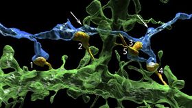
Video showing all of the separate components that make up the tiny section of mouse brain. Credit: Kasthuri/Guardian Science and Tech/YouTube.
The researchers decided to focus on the region of the mouse brain that receives the sensory signals coming from the whiskers. By taking thousands of minuscule slices just 29 nanometers thick, the trick was to be able to use an electron microscope to automatically take photos of each slice, giving them a resolution so fine that they could even distinguish between individual molecules. The photos were then stitched together to form the stunning 3D images. For context purposes, one pixel on an MRI is around one billion pixels in these images, Kasthuri told The Guardian.
The section in the video is just a tiny portion of the mouse brain for which they reconstructed all the connectivity, in fact it measures just 1.5 mm3. The task of mapping the entire brain would be monumental, not only taking an estimated five years, but generating billions of gigabytes of data. The researchers hope that they can image the brains of people with physiological conditions, such as schizophrenia, and compare them with neurotypical brains to find out how the connections differ.