

Mankind’s battle with cancer has been raging for thousands of years, and while the addition of new treatment techniques to our medicinal arsenal may soon give us the upper hand, researchers still believe that our chances of victory can be improved by learning more about how cancer has affected us throughout history.
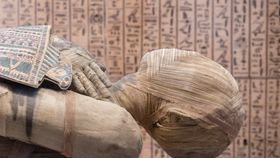
Mummies, such as those dating back to Ancient Egypt, are therefore a hugely valuable treasure trove of information, although because scientists aren’t sure what a mummified tumor looks like, locating cancerous cells in these ancient bodies has so far proved quite challenging.
To try and solve this conundrum, a doctoral student at Western University in Canada named Jennifer Willoughby has been mummifying cancerous mice – donated by a cancer lab – in order to figure out exactly what mummified tumors look like.
Presenting her wrinkled rodents at the World Congress on Mummy Studies in Lima, Willoughby revealed how she used a range of different preservation techniques. Some of the mice were buried in sand in a hot terrarium, to mimic the natural mummification process that some bodies undergo. Others, meanwhile, were given the full pharaoh treatment, with Willoughby carefully removing their internal organs – minus their brains, which couldn’t fit through their tiny noses – before filling their abdomens with a dehydrating agent called natron, originally used by the Ancient Egyptians.
After letting their tiny bodies dry for 50 days, Willoughby then dipped the mice in pine resin, wrapped them in bandages and coated them in beeswax, before anointing them with frankincense and myrrh, and finishing the ritual with an ancient prayer.
With the fun part over, the bioarchaeology student placed the mouse mummies in a tomography scanner, discovering that the tumors were indeed distinguishable from other soft tissues, appearing more solid and interacting with X-rays in a particular way.
Using this knowledge, researchers should now know what to look for when seeking out mummified tumors, which could lead to new insights into how humans have been affected by cancer in the past.
[H/T: Science]