

When suspected cancerous tissue is found within a patient’s brain during surgery, there is simply no time to remove a sample, send it to a separate laboratory, and wait for a diagnosis following microscopic analysis. A study in the journal Biomedical Optics Express describes a new handheld microscope that has the ability to image various tissues with as much detail as the best laboratory equivalents. This could potentially allow surgeons to look for miniature, malignant features during operations – including perhaps neurosurgery.
To confirm the presence of a brain tumor, a neurosurgeon will open up the skull and carefully delve inside. The choice then has to be made to remove the potentially malignant tumor or, if it appears to be benign, leave it. Removing it carries the risk of damaging healthy neurological tissue, but leaving it – even a part of it – means that it may potentially continue to develop into a metastasizing, untreatable tumor.
Researchers at the University of Washington (UW) are currently engineering a miniature, powerful microscope that could solve this problem. It uses two different optical technologies that may give brain surgeons a real-time assessment of whether or not a suspected tumor in the brain is truly cancerous.
“Surgeons don't have a very good way of knowing when they're done cutting out a tumor,” said senior study author Jonathan Liu, UW assistant professor of mechanical engineering, in a statement. “They're using their sense of sight, their sense of touch, pre-operative images of the brain – and oftentimes it's pretty subjective.”
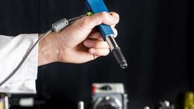
The pen-sized microscope he is helping to develop aims to bring more objectivity into the operating room. Using an approach called “dual-axis confocal microscopy,” it is able to brightly illuminate and peer through very thin sheets of tissue. It can capture details of up to half a millimeter; at this scale, some types of cancerous cells can be seen.

Real-time images produced by the new device (black and white) show similar detail to conventional pathology laboratory microscopes and imaging techniques (color). (a-b) Tongue tissue; (c-f) Kidney tissue; (g-h) Colon tissue. Yin et al./Biomedical Optics Express
Light scattered in multiple directions by the target tissue produces erroneous “background noise” for conventional microscopes. This dual-axis set up is able to detect and reject this background noise, producing more detailed images.
In addition to this, a technique called line scanning is employed by the microscope to speed up the image processing. Specialized reflecting surfaces known as micro-electrical-mechanical (MEMS) mirrors direct the microscope’s optical beam with remarkable precision, allowing it to rapidly scan the tissue, piece by piece, in order to build up a high-resolution image in record time.
Impressively, the increased portability was possible without compromising on image detail. In the paper, using mouse tissue the researchers demonstrated that subcellular features could be picked up at resolutions comparable to the clunkier, desk-based models used in pathology laboratories. Blood cells being trafficked within capillaries were also shown to be precisely tracked by the device.
While the team has not tested its capabilities on tumor cells, if shown to be effective at identifying cancerous tissue in mice and human brains during trials, it is hoped that the tool will be used in the clinic within the next four years.