


Trialling the technology for the first time in Europe, surgeons in London have used a new laser technique to identify abnormal tissue during an operation to remove a brain tumor. The method allows the surgeons to determine which material needs to be excised in the operating theater, rather than sending the sample off for analysis during surgery.
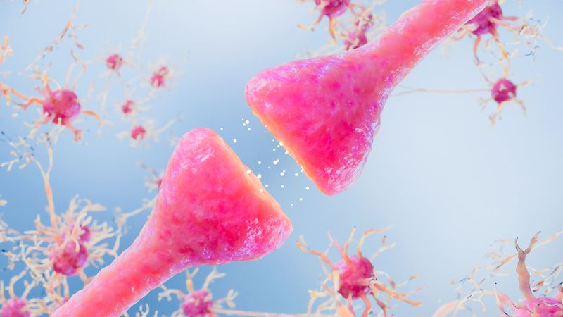
The new piece of kit works by firing a near-infrared laser into the exposed brain during the surgery to remove the already diagnosed tumor. The beam of light from the laser causes molecules in the cells to vibrate. The fibre optics attached to the laser pick up the scattered light that is reflected and, because the molecules in the healthy tissue vibrate at a different frequency to the abnormal tissue, the tech is able to distinguish between these two signatures. This allows the surgeon to know if they have removed all of the tumor instantly.
Normally, the surgeon will remove small samples of the brain and send them off for analysis mid-operation, but this can take up to 40 minutes to get the results back. This new system takes only around a second to get a reading, and can buy the surgeons valuable time. “Optical technologies like this are the future. They are fast and don't destroy any tissue and could be used during many types of cancer surgery or when dealing with infection like a brain abscess,” neurosurgeon Babar Vaqas explained to BBC News.
In addition to this, the operation, which took place Charing Cross Hospital, also demonstrated the amazing potential of what is being called the “iknife.” This is an electrosurgical knife that works by using an electrical current to rapidly heat the tissue, cutting through it in a way that reduces blood loss and produces smoke at the same time. While this technique was invented in the 1920s, researchers from Imperial College London realized that the smoke the knife produces could hold vital biological information.
By linking the electrosurgical knife up to a mass spectrometer, which is used to identify what chemicals are present in a sample, they were able to read the information found in this smoke. “This is bringing the laboratory into theater, giving a real-time molecular fingerprint of tissue,” Kevin O’Neill, head of neurosurgery at Imperial told BBC News. “The potential is amazing, not just to differentiate between normal brain and tumor, but whether the patient is likely to respond to specific treatments.”
The operation in which they used these two new pieces of tech was filmed for the BBC, and you can watch how it went here. During the surgery, the patient Reuben Hill was asked to sing to make sure no damage was done to the speech centers as they removed the tumor in his brain. The surgery was a complete success, and Hill has since found out that the tumor was non-cancerous.
[H/T: BBC News]