

If you’ve ever broken a bone, chances are you’ve seen one of the standard black-and-white X-ray images that physicians have been using to diagnose internal injuries for decades. But this technique – and even the more recent modalities like CT and MRI scans – can provide only a limited glimpse of the state of tissues and structures underneath our skin.
A new platform developed by imaging researchers at CERN could change all that.
The breakthrough Medipix3 scanner creates high-resolution, 3D, color images of living structures. Born from technology designed to track particles in the Large Hadron Collider, the Medipix3 works by emitting a targeted beam of electromagnetic radiation at the frequency of X rays at the object of interest, then detecting, counting, and differentiating every particle, or photon, that hits its sensor on the other side – sort of like a souped-up version of a typical digital camera.
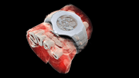
Next, the device’s advanced algorithms interpret the spectroscopic information and convert it into images. The high sensitivity and accuracy of the scanner allows for an unprecedented visualization of all the varied substances within the human body, such as fat, liquids, and different minerals. And as demonstrated by the image with the wristwatch, the platform can also easily differentiate metals.

In contrast, a traditional X-ray scanner works by emitting a wave of X-ray radiation at a biological structure and detecting the particles that pass through using light-sensitive film or a sensor. Since the rays pass through less dense materials (like fluid and fat) more easily and lose less energy in doing so, the image created on the other side is much like a shadow of the heavier structures in your body. But these old-school sensors and film are not very good at differentiating the energy levels of the particles that strike them, resulting in a low-resolution image that fails to capture the numerous types of similarly dense tissue and subtle variations and abnormalities that could represent disease.

According to CERN, the father and son team of Phil and Anthony Butler, also based out of Canterbury and Otago Universities, spent a decade tinkering and refining the technology from earlier generation particle-tracking platforms built by the nuclear research hub and collaborations between more than 20 other research institutions.
The Butlers' company, MARS Bioimaging, was launched to bring the innovations developed by this diverse group of leading minds to the commercial market, thus enabling hospitals and clinics to begin using it.
Several research groups have already used a small version of the scanner in investigations of a variety of health conditions, to great success.
“In all of these studies, promising early results suggest that when spectral imaging is routinely used in clinics it will enable more accurate diagnosis and personalisation of treatment,” Anthony Butler said in the CERN statement.
Though widespread availability is probably still years away, the developers are excited about the variety of applications beyond simple bone fracture imaging that the system presents, including visualization of fatty plagues within vessels, monitoring nanoparticle drug delivery systems, observing the changes to the calcium lattice of bones in people with osteoporosis, and examining how medical implants are integrating with surrounding tissue.