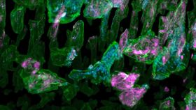


Researchers have captured footage of cells growing, changing, and moving within a live animal at the highest resolution yet. The team combined two imaging techniques to create a detailed 3D movie that shows how cancer cells, spinal nerves, and immune system cells behave in the inner ear of a developing zebrafish.
The rest of this article is behind a paywall. Please sign in or subscribe to access the full content.The new approach, published in Science, addresses several concerns scientists had about previous approaches to cell microscopy. Cells are either extracted and analyzed in vitro, like on little glass slides, or in vivo, which is directly in the animal but uses such intense light that it may change their properties.
"This raises the nagging doubt that we are not seeing cells in their native state, happily ensconced in the organism in which they evolved,” senior author and Nobel prize winner Eric Betzig, from the Howard Hughes Medical Institute, said in a statement.
"It's often said that seeing is believing, but when it comes to cell biology, I think the more appropriate question is, 'When can we believe what we see?'"
For one movie, the team was able to observe an orange immune cell interacting with blue sugar particles. Another video shows a cancer cell trying to attach itself to the wall of a blood vessel. This is an incredible view of the microcosmos that live inside organisms.
The new approach uses lattice light sheet microscopy, which uses thin sheets of light to rapidly but gently capture 2D images. These can then be used to construct a 3D image. Betzig and his team paired this with adaptive optics, a technology used in astronomy that helps astronomers get clearer images of distant objects.
While these technologies had already shown promise in microscopy, they hadn’t been employed together and, due to their cost, many thought they weren’t worth the effort. The new research demonstrates that they can indeed produce incredible results. Still, there’s more to do.
"Technical demonstrations and publications don't amount to a hill of beans. The only metric by which a microscope should be judged is how many people use it, and the significance of what they discover with it," Betzig added.
The microscope takes up a 3-meter (10-foot) table, so it’s not exactly portable technology. The team are now working to make it smaller and more affordable so that individual labs can use it.