

An image is said to tell a thousand words. Well, now there is an electron microscope that can tell us 1 million. Researchers have developed a new approach to electron microscopy that not only allows us to see individual atoms but to also learn about several of their properties at the same time.
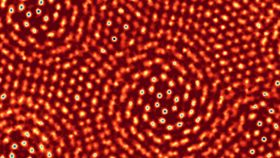
The technology is called an electron microscope pixel array detector (EMPAD). The tech, developed last year, goes beyond imaging individual atoms – it was employed to study two one-atom-thick layers of molybdenum disulfide, with one layer on top of the other and slightly askew so that the single atoms are visible. EMPAD reached a world-record resolution for such an image.
As reported in Nature, researchers were able to resolve a distance of 0.039 nanometers, a distance smaller than the smallest atom. The usual distance for atomic bonds is 0.1-0.2 nanometers, with the EMPAD able to visualize these bonds.
"It's essentially the world's smallest ruler," co-author Professor Sol Gruner, from Cornell University, said in a statement.
The resolution of the microscope was so good, even at low energy, that the team was able to discover that within the lattice of atoms there was a single sulfur atom missing from one of the molybdenum disulfide layers. “A defect in the lattice,” Gruner explain – in a 2D material. “That’s astounding to me.”
EMPAD has been retrofitted on many different microscopes across the Cornell University campus and tested with many different intensities. Electron microscopes see electrons, just like regular cameras see photons (light particles). The ability of EMPAD to detect not only the direction but also the speed of the incoming electrons allows for incredibly high-resolution. The tech has been tested successfully with intense beams containing up to a million electrons.
“The analogy I like to use is, a car is coming at you at night,” Gruner added. “And you’re looking at the lights coming at you, and you’re able to read the license plate between them without being blinded.”
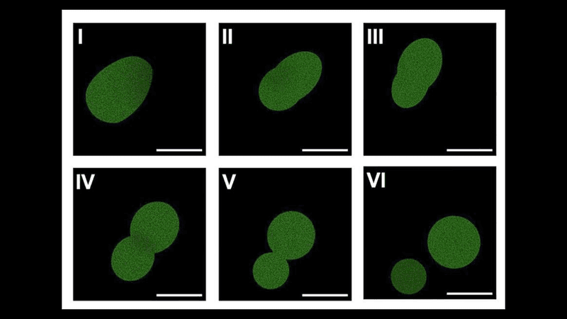
The team believe that EMPAD can be used successfully in living cells. Since the energy of the electron beam is lower than what is often used in electron microscopy, it could be used to look at cellular processes without damaging cells.