

A new project is giving a never-before-seen look into the beautiful and, let’s face it, bizarre world of human embryos. Not only could it help doctors understand congenital malformations and diseases better, it’s showing how surprisingly little modern science knows about the physiology of early human embryo development.
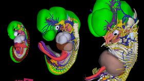
The rest of this article is behind a paywall. Please sign in or subscribe to access the full content.Embryology experts from the University of Amsterdam have developed interactive three-dimensional digital models of embryos. The so-called “3D Atlas of Human Embryology” is part of a study recently published in the journal Science
"It's fair to say that we currently know more about the Moon than about our own embryonic development,” researcher Bernadette de Bakker, of the University of Amsterdam, told IBTimes UK.
"The current textbooks all show the same kind of images based on embryonic specimens from the 1930s. Subsequent books kept using the image, only updating new molecular information," she added.
The atlas was developed with the aid of 75 college students, who analyzed around 15,000 stained samples of tissue sections, some of which date from 100 years ago, collected from the Carnegie Collection of human embryonic specimens.
The result is 14 interactive models that detail the human embryo at different stages within the first 60 days of development. It also allows you to flick through different systems of the body as they develop and come into shape, including the respiratory system, the skeletal system, nervous system, and many others.
Speaking to the Guardian, de Bakker added that many illustrations of human embryos used in medical training still use studies of mouse and chick embryo development, even today.
“We discovered that some organs in humans develop earlier than they first arrive in chick or mouse embryos and some [develop far] later…. It is a beautifully careful assessment of development through analysis of material with limited availability. It will provide an invaluable atlas to guide future study,” she said.
You can check out the digital models for yourself on the 3D Atlas of Human Embryology website, available on Microsoft Windows and Mac if you open it using either Adobe Acrobat or Acrobat Reader. (Note: it doesn’t work as well if you read it in the web browser, so it's best to download it)