

In the New Kingdom of Ancient Egypt, after the death of a pharaoh, embalmers would remove the brain matter of their ex-ruler via the nose. Now, we’ve improved on both counts: surgeons can go in through a keyhole incision by the eye socket, remove a brain tumor, and the patient doesn't just survive but is much better off for it
The rest of this article is behind a paywall. Please sign in or subscribe to access the full content.“I was amazed by the recovery,” Ruvimbo Kaviya – a 40-year-old woman from the UK who, in February last year, became the first person in the country to undergo endoscopic trans-orbital surgery to remove a tumor – said in a statement. “I was only in the hospital for two days, with no side effects or swelling. I feel perfectly fine now.”
After months of increasingly intense headaches, eventually developing into spasm-like sensations she initially thought were due to toothache, in 2023, Kaviya was eventually diagnosed with meningioma – a type of tumor that grows in the protective layers of the brain known as the meninges. An MRI revealed it was located in the cavernous sinus – a hollow space found behind the eye socket, through which the jugular vein carries blood away from the brain.
That would, in times past, be a problem. The cavernous sinus is hard to access, and “considered somewhat inoperable”, per the NHS statement. But Kaviya’s diagnosis came at a fortuitous point, as in recent years, surgeons have been experimenting with less-invasive procedures to remove such tumors – which would previously have required major incisions, the removal of parts of the skull, and long recovery times.
“This technique allows us to remove tumors without opening the skull or having to retract or compress the brain,” explained Asim Sheikh, Consultant Skullbase and Neurovascular Neurosurgeon at Leeds Teaching Hospitals NHS Trust, and a member of the team who carried out the procedure – all necessary steps in traditional methods to reach the cavernous sinus.
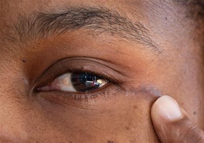
But the new technique avoids the brain almost entirely, “significantly reduc[ing] trauma” and “enabling patients to recover faster with minimal visible scarring,” Sheikh said. Surgeons instead accessed the tumor via a tiny incision next to Kaviya’s eyelid – leaving just a 1.5-centimeter (0.6 -nch) scar.
A 3D model, courtesy of the Trust’s 3D Planning Service, helped the procedure succeed – a technology that Jiten Parmar, Consultant in Maxillofacial Surgery at Leeds Teaching Hospitals NHS Trust, described as “a game changer”. It allowed the team to do practice runs of the surgery, performing steps on a life-size model of Kaviya’s brain and skull to perfect their technique before the big day.
“When the surgical team approached me, we used scans of Ruvimbo’s brain and skull to create a 3D replica model,” explained Lisa Ferrie, the Trust’s Biomedical Engineer and 3D Planning Service Lead.
“This technology enabled the team to study her anatomy in detail and prepare for the procedure with unparalleled accuracy,” Ferrie said. “Seeing the model and knowing it contributed to this groundbreaking surgery is incredibly rewarding.”
For Kaviya, though, being the first in the UK to undergo the procedure was never the main goal. “It was the first time they were doing the procedure,” she is reported to have said, but “I didn't even think about [that].”
“I had no option but to agree because the pain was just too much,” she said. “All I needed was for it to be removed.”