

A team of tissue engineers and plastic surgeons in China have created new ears for five children using a combination of autologous cell culturing and 3D printing.
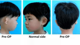
The first patient, a girl who was 6 years old when the process began, now has a realistic-looking ear and suffers no serious side effects 2.5 years after implantation.
She and the other four patients were all born with unilateral microtia – a congenital deformity that results in an undersized and malformed outer ear. The current treatment option for microtia involves harvesting cartilage from the patient’s ribs and carefully shaping the tissue into an ear shape.
Unfortunately, this method “inevitably leads to donor site injury, and replicating the complex 3D ear structure is hard to achieve using surgeons' hand skill,” according to the group’s paper, which is published in EBioMedicine.

Following the 1997 breakthrough of the “Vacanti mouse”, medical researchers have been experimenting with using the body’s own cells, combined with a structural scaffold, to create refined and functional replacements for human organs. The pioneering technique described represents the first time human ear-shaped cartilage has been grown in vitro (outside a living body) and then surgically implanted.
To begin, the group took detailed CT scans of each patient’s healthy ear. They then used design software to mirror the images and convert the shape into a 3D-printed mold. Next, the mold was cast with a porous, biodegradable material called PGA. A more rigid material called PCL was used to reinforce the core of the scaffold.
With a polymer "skeleton" of the patient’s desired ear ready to go, cartilage-producing chondrocyte cells were isolated from the malformed ear tissue. After the cells had multiplied sufficiently, they were spread into the molds and incubated with a steady diet of growth factors. During this 12-week process, the chondrocytes began to form collagen and elastin fibers within the spongy PGA lattice.

As the cell matrix expanded, the PGA was slowly degrading; By the time the engineered ear was ready for implantation, it was primarily composed of the child’s native tissue and only small amounts of artificial material remained.
Meanwhile, a tissue expander was slowly stretching the skin over the extraction site in order to make room for the large implant. Once ready, the cultured cartilage was carefully placed in the skin pocket by plastic surgeons.
Over the next 2.5 years, patient one was monitored and underwent several cosmetic surgery adjustments. Tiny tissue samples removed during these procedures proved that the chondrocytes remained healthy and continued to produce cartilage comparable to that of a natural ear.
Moreover, the only artificial substance remaining in the engineered ear was part of the PCL core – as intended. Previous attempts to implant cultured ears have failed because the tissue used was not rigid enough to maintain its shape against the physical assaults of daily life. The Chinese team’s ear appears to be holding up well, although they note that its long-term integrity – after the PCL core fully degrades by year four – remains unknown.
Unfortunately, the next four patients showed less consistent results. One child’s new ear failed to produce new cartilage and the others are less refined aesthetically. All patients will be monitored for up to five years post-implantation.

Though the technique clearly needs to be perfected further, these early results are a promising leap forward for the field of reconstructive medicine.
“These are the steps we need to make to bring this technology to patients,” said Jos Malda, a biofabrication and regenerative medicine professor at Utrecht University, to New Scientist. “It’s quite an achievement.”