

The human brain’s waste clearance system has been visualized for the first time ever, confirming that the complex organ does indeed contain its own lymphatic vessels.
The rest of this article is behind a paywall. Please sign in or subscribe to access the full content.Previously, scientists were unsure how the brain disposed of its metabolic by-products, but a revolutionary new scanning technique has finally provided a glimpse of the cerebral lymphatic system in action.
As the seat of cognition and the control room for the body’s functions, the brain has some considerable energy demands and is unsurprisingly the most metabolically active organ. This means it also produces an awful lot of waste, which must be efficiently cleared to prevent a build-up.
Throughout the body, waste is filtered through the lymphatic system – yet researchers had never previously managed to scan the structures that facilitate this process in the brain. Among other challenges, visualizing this process via magnetic resonance imaging (MRI) would require the injection of potentially dangerous contrast agents, making it impossible to apply this method to live subjects.
Due to a lack of direct evidence for the presence of lymphatic vessels in the brain, it was previously believed that waste products were cleared from the cerebrospinal fluid (CSF) via blood vessels. However, recent studies on rodents have indicated the presence of meningeal lymphatic vessels, raising the possibility that similar structures may also conduct the clearance of waste from the human brain.
For a new study in the journal Nature Communications, researchers developed a safe, non-invasive MRI protocol that bypasses the need for toxic contrast agents. Instead, their technique produces images based on the natural contrast gradient that exists between the protein-rich contents of lymphatic vessels and the more diluted CSF.
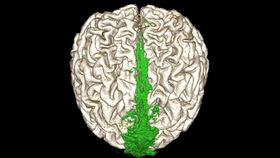
Using this method, the authors scanned the brains of 81 epilepsy patients, revealing the presence of lymphatic structures running parallel to both the venous sinuses and the cranial nerve. These vessels direct the flow of waste products out of the skull, connecting to lymph nodes in the neck.
“This is the first report to show the complete human brain lymphatic system architecture in living humans,” explained study author Onder Albayram in a statement.
MRI showing the dorsal flow of the brain’s waste clearance system (shown in green).
Importantly, rodent studies have indicated that the functioning of this meningeal lymphatic system begins to decrease in old age, and some researchers have speculated that this may contribute to pathologies such as Alzheimer’s disease and age-related cognitive decline.
With participants in this latest study varying in age from 15 to 80 years, the authors took advantage of the opportunity to observe how the human brain’s lymphatic structures differ between older and younger subjects.
Their analysis indicated that the thickness of meningeal lymphatic vessels increases with age, resulting in a reduction in their capacity to remove waste.
Based on this discovery, the authors conclude that their non-invasive imaging technique “may allow for new approaches in diagnosis or treatment of neurological disorders such as traumatic brain injury, Alzheimer disease, [and] multiple sclerosis.”
According to Albayram, the new imaging method could also lead to new understandings of how other challenges affect the brain’s capacity to function. For example, “what happens during a TBI [traumatic brain injury]? Are the lymphatic vessels damaged, and how do they recover? This technique will enable us to begin to answer these questions.”