Health and Medicine
 PUBLISHED


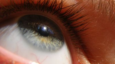


A new device that pumps oxygenated fluid through donated eyeballs significantly improves their longevity outside the body and even enables pig eyeballs to transmit electrical signals over 10 hours after death.
The device, called the Eye-in-a-Care-Box (ECaBox), is 3D-printed and works by circulating an oxygenated solution around the eyeball, pumping it into the eye through a flexible tube inserted into the main artery and using sensors to regulate flow and pressure levels.
A transplant of a working eye has never before been achieved in humans, despite our success with other organs including the heart, lungs, and kidneys. One reason for this is that the retina – the tissue at the back of the eye that converts light into signals that can be transmitted to the brain – has a delicate structure that is extremely vulnerable to even temporary losses in oxygen.
In 2024, a pioneering surgery successfully transplanted part of a face and also a new left eye into a man who had received an extreme electrical shock. While he didn't regain sight in the transplanted eye, researchers showed that his new retina responded to light signals and had regained blood flow in a process called reperfusion.
Following that success, researchers led by a team at the Barcelona Institute of Science and Technology in Spain saw the potential of perfusing the eye outside of the body, or ex vivo, in order to prolong the life of the organ and improve chances of one day being able to perform a successful transplant of a working eye. Their results have now been posted to an online preprint server, though they have yet to be peer-reviewed.
The team first tested their device with pig eyes obtained from a nearby slaughterhouse, which could be extracted, put on ice, and transported to the laboratory within 2.5 hours post-mortem. When they put some of those eyes in the ECaBox and left others outside, they found that the eyes in the box were "significantly more viable" after 24 hours compared with those that had been stored outside at 4°C (39°F).
The ECaBox comes with a window on the side that allowed the researchers to monitor blood flow in the reperfused eyes using a coloured dye. They saw liquid flowing through vessels in 90 percent of well-cannulated eyes, and when they dissected them and put them under a microscope they could confirm they had perfused even at the level of capillaries and micro blood vessels.
In addition, by using a technique called electroretinography (ERG) – which measures the electrical response the retina produces when it's hit by light – the team found that 15 out of 36 perfused pig eyes still generated light responses in the device, in some cases for up to 10 hours after death. When they switched the perfusion off, the signal faded – a strong sign that it was the oxygenated flow keeping the retina alive and responsive.
From their experiments in the pig eyes, the researchers concluded that vessels in eyes that received perfusion more than 5 hours post-mortem lost integrity in a way that was "statistically significant" compared with those perfused at earlier times.
The researchers also carried out some experiments on 12 human eyes from six post-mortem donors. They divided these up so that one eye from each pair went into an ECaBox and one was left outside. As with the pig eyes, they found that the perfused eyes fared much better than their counterparts.
That said, the researchers did not report ERG recordings from the human donor eyes, so it remains unknown whether they retained light responses similar to those seen in the pig experiments. Because the human eyes arrived 6 to 10 hours after death, they may have been less likely to retain retinal function, but this was not tested.
The number of human donor eyes in the experiment is also rather small, and the researchers put this down to ethical and practical difficulties. Donor eyes are generally used for corneal donation, with the cornea, the clear front part of the eye, being transplanted into patients.
The team hopes that the potential of this work might provide enough of a case to give them access to more and better-quality donor eyes in future, and they have been designing a portable surgery room to help get around the practical limitations of perfusing an eye in the smallest amount of time post-mortem as possible.
All that said, keeping the retinal tissue functional is not the only thing preventing working eye transplants from being viable. Even if the retina is producing signals, these won't go anywhere unless the optic nerve can be wired back up to the transplant-recipient's own brain.
"Preservation of eyes for transplantation is potentially helpful," James Bainbridge at University College London, who was not involved in this work, told IFLScience, "[But] restoration of sight will depend on rewiring of the optic nerve, and this challenge has yet to be addressed.
One way ECaBox might be useful in the short term is as a platform for studying the pathology and treatment of degenerative diseases, write the researchers.
While it's possible to study living eyes in mice, they lack a key structure called the macula, which is central to human vision and is the primary site of damage in diseases such as age-related macular degeneration.
Larger animals have this structure, but they are practically and ethically more difficult to work with. If their eyes could be donated after death and kept functional in an ECaBox, it might open up a whole slew of possible experiments.
The preprint is available on BioRxiv.