

3D printing is slowly becoming invaluable in medicine. Not only can scientists make 3D-printed body parts, but they can also use the technology to help guide life-saving surgery. Just last week, for example, we heard the story of a team practicing brain surgery using a 3D replica of a woman’s entire brain vasculature. And now, the technology has triumphed again, helping doctors separate conjoined twins.
Born on April 11 of last year, twins Adeline and Knatalye Mata from Lubock, Texas were extensively fused, from the chest all the way down to the pelvis. This presented surgeons with an extremely convoluted separation task, perhaps one of the most intricate to date.
Knowing how complex the procedure would be, rather than relying solely on scans to guide the knives, the team decided to use these images to create replicas of the relevant body parts. After CT scans were taken of the twins, the physicians could see that the hearts were not joined, and while the livers were, there appeared to be a plane of separation that could potentially be sliced though.
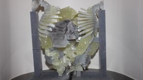
The images were then used to fabricate both skeletal structures and organs using multi-material 3D printing. For the former, a hard plastic resin was used, while the softer organs were printed using a rubber-like material. The team also printed the livers separately, using a white resin to highlight the major vessels. The replicas were also printed in such a way that they could be assembled or detached as the team prepared for the real procedure.
Image credit: CT scan data alongside 3D-printed replica. Krishnamurthy/Radiological Society of North America
Ten months after the twins were born, a team of more than 26 physicians, 12 of which were surgeons, assembled to undertake the momentous separation task, which in total took more than a day to complete. Much to the group’s relief, the models turned out to be a remarkably accurate representation of the actual anatomy of the infants, thus their preparations and simulations based on these replicas were not fruitless.
“The surgeons found the landmarks for the liver, hearts and pelvic organs just like we had described,” study lead author and chief of radiology research and cardiac imaging at Texas Children’s Hospital, Rajesh Krishnamurthy, said in a statement. “The concordance was almost perfect.”
Although there was one bleeding complication during the procedure, which wasn’t due to a discrepancy between the models and the twins’ anatomies, the surgery was a success, with both twins returning home early this summer.
It’s clear such a use of 3D printing has a place in the operating theater, but whether it will become a standard procedure for extremely complex surgeries is uncertain at this stage. Unfortunately, in the U.S. at least, what seems to be holding it back is money.
“The 3D printing technology has advanced quite a bit, and the costs are declining. What’s limiting it is a lack of reimbursement for these services,” explained Krishnamurthy in the statement. “The procedure is not currently recognized by insurance companies, so right now hospitals are supporting the costs.”