

Cytotoxic T cells, which researchers describe as ‘serial killers,’ have a pretty important role to play in keeping your body healthy. They move rapidly around their environment looking for infected and cancerous cells. Once identified, cytotoxic T cells lock on to their target and kill them. This remarkable process has now been captured on film by researchers from the University of Cambridge using state-of-the-art imaging techniques.
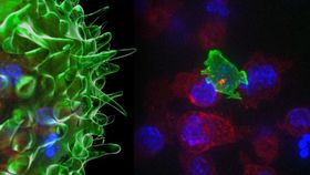
There are billions of cytotoxic T cells in our body, which are the orange or green 'blobs’ in the video below, and they are able to recognize a variety of pathogens through the ‘markers’ on the surface of the cells. These markers, known as antigens, tell the cytotoxic T cells whether the cell is carrying foreign or abnormal molecules. Once cytotoxic T cells recognize unwanted intruders, they launch an attack by binding to the cell and injecting it with poisonous molecules called cytotoxins—shown in red in the film.
"In our bodies, where cells are packed together, it's essential that the T cell focuses the lethal hit on its target, otherwise it will cause collateral damage to neighboring, healthy cells," says Professor Griffiths. "Once the cytotoxins are injected into the cancer cells, its fate is sealed and we can watch as it withers and dies. The T cell then moves on, hungry to find another victim."
Though cytotoxic T cells are tiny—measuring a tenth of the width of a human hair—they are a vital line of defense against infections and tumors. The study, published in the journal Immunity, used high-resolution 3D time-lapse multi-color imaging to record these events onto films. Using the live-cell imaging techniques called spinning disk confocal microscopy and lattice light-sheet microscopy, researchers were able create 3D images by ‘stitching’ together captured slices.
You can watch the epic battle below.