

Thanks to fresh data and a cutting-edge computer modeling tool, you can now take the most sophisticated look yet at SARS-CoV-2, the minuscule bundle of proteins and lipids that has managed to infect at least 50 million people worldwide so far.
The rest of this article is behind a paywall. Please sign in or subscribe to access the full content.Scientists have developed a new three-dimensional rendering of SARS-CoV-2, the coronavirus behind the Covid-19 pandemic, using the latest microscope data and scientific speculation. Details of their modeling tool, which can be updated to reflect the latest data at any time, are published in the journal IEEE Transactions on Visualization and Computer Graphics.
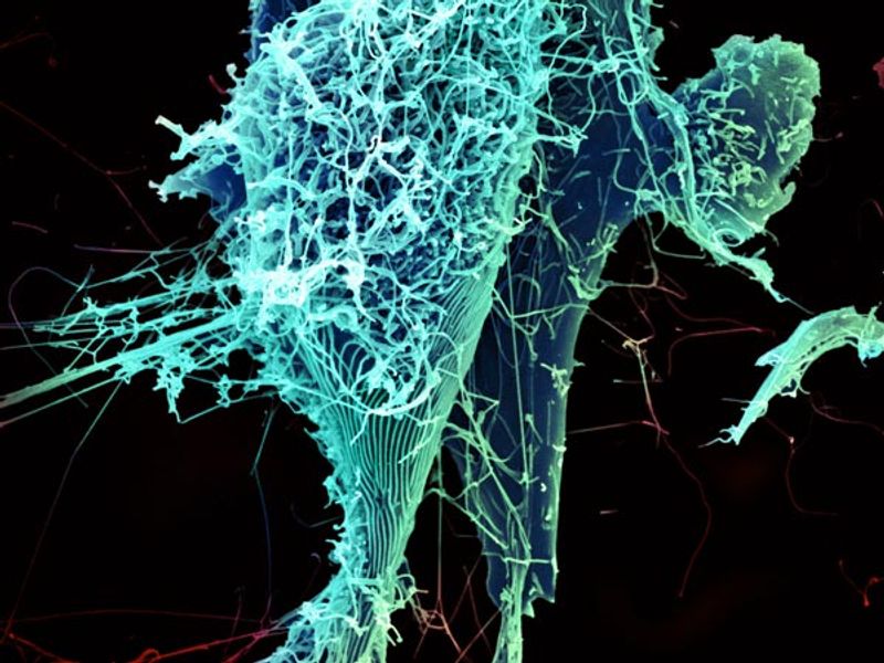
Viruses are absolutely tiny, with a generic coronavirus measuring between 120 to 160 nanometers in diameter. That’s hundreds of times smaller than the average bacteria or human red blood cell. As such, visualizations of viruses can often miss out on detail and accuracy. Using their recently developed modeling tool, however, researchers from the King Abdullah University of Science and Technology (KAUST) in Saudi Arabia were able to visualize components in the virus down to just 10 to 100 nanometers in size.
"Our 3D model demonstrates the current state-of-the-art structure of SARS-CoV-2 at the atomistic level and reveals details of the virus that were previously impossible to see, like how we think nucleocapsid proteins form a rope-like structure inside it," Ondrej Strnad, study author and research scientist from KAUST, said in a statement.
Coronaviruses are infamous for their spike protein, the major surface protein that they use to bind to cells and invade. The name "coronavirus" is derived from the Latin word “corona,” which means "crown,” making reference to these crown-like spikes that coat the surface. This new visualization goes a step further than just showing the viruses' surface and attempts to even map the lesser-known innards of the virus.
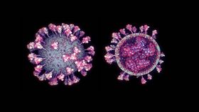
"Our model shows the complete viral ultrastructure as we know it to date, and not just some arbitrarily placed, incomplete spike proteins on a lipid membrane," said Ngan Nguyen, lead study author and computer science PhD student at KAUST.
"Other available models also don't show the interior of the virus, as its details are not currently known. Scripps Research Institute US provided us with the most likely hypothesis for the structure's interior based on current data. If this hypothesis is proven wrong, then we can easily update the model," Nguyen continued.
The new project is impressive, but not just a pretty picture. The researchers hope their work could be used by other researchers to better understand the virus and find ways to tackle the ongoing outbreak. For example, figuring out the shape of the spike protein is one of the major clues to finding drugs that can target the virus and block it from entering our cells.
For more scientific glimpses of SARS-CoV-2, check out this other visualization created by award-winning biomedical visualization studio Visual Science earlier this year.