

The rest of this article is behind a paywall. Please sign in or subscribe to access the full content.
Doctors have removed a tumor that contained several fully formed teeth from the brain of a 4-month-old male infant from West Virginia. The procedure was described (and visualized in graphic detail) in the New England Journal of Medicine last year.
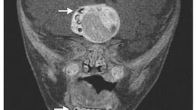
When the circumference of the baby’s head appeared to be increasing during his routine pediatric visits back in 2012, doctors used ultrasound, CT, and MRI to scan his brain. These images revealed what University of Maryland’s Narlin Beaty and Edward Ahn of Johns Hopkins Children’s Center call “heterogeneous, enhancing suprasellar mass.” Its dimensions were 4.1 by 4.0 by 3.5 centimeters. Then they noticed multiple structures along the right side of the mass that seemed to resemble teeth that are normally found in the lower jaw.
 When the child underwent brain surgery, multiple fully-formed teeth were taken out along the tumor mass. After examining the pathological tissue, researchers revealed that the tumor was an adamantinomatous craniopharyngioma. These rare, slow-growing tumors arise from Rathke's pouch, an embryonic precursor to the anterior pituitary -- one of the lobes of the pituitary gland at the base of the brain; this pea-sized gland releases many of our important hormones.
Previous work has suggested that craniopharyngiomas come from cells that also form teeth. "No one knows," Ahn told the Baltimore Sun. "It must be the origin of the tumor… They might have had some common lineage with the cells that produce teeth." And while these tumors do have calcium and keratin inside, they’re not usually organized as teeth.
"It's not every day you see teeth in any type of tumor in the brain. In a craniopharyngioma, it's unheard of," Beaty told Live Science. Calcium deposits, even in the form of flakes, are common in these types of tumors, "but when we pulled out a full tooth… I think that’s something slightly different." In the past, when teeth have been found in human brains, these involved tumors called teratomas. Unlike craniopharyngiomas, which only have one layer of tissue, teratomas contain all three tissue types found in early human embryos. That’s why they have been known to contain fingers and partially formed humans.
As of a year ago, the patient is making good progress.
Images: Narlin Bennet Beaty, M.D., and Edward Ahn, M.D., NEJM 2014