

The dramatic rise in cases of children being born in Brazil and other parts of South America with underdeveloped brains led to the World Health Organization declaring the Zika virus a global public health emergency. While it is highly suspected that the virus is causing children to be born with microcephaly, the link is difficult to definitively prove. Yet the increase in cases and its rapid spread has meant researchers have been working flat out to try and pin this link down, as well as find some way to stop the virus in its tracks.
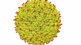
Now scientists from Perdue University have made a leap forward in this quest for a cure, publishing the first ever atomic-scale view of the structure of the virus. “The structure of the virus provides a map that shows potential regions of the virus that could be targeted by a therapeutic treatment, used to create an effective vaccine or to improve our ability to diagnose and distinguish Zika infection from that of other related viruses,” explains Richard Kuhn, the leader of the research team who figured out the structure.

The virus has been linked to a massive rise in microcephaly cases in South America. Mario Tama/Getty
Using a technique known as cryo-electron microscopy allowed the researchers to image the Zika virus in a matter of months rather than the years it would have taken with the more traditional X-ray crystallography. By first freezing the virus, they then fired electrons at it, creating thousands of 2D images that they then stitched together to form a detailed 3D composite image of the virus. And what it shows is quite revealing.
The overall structure turns out to be incredibly similar to other flaviviruses, a group that contains other more well-known viruses such as dengue and yellow fever. It has wrapped its RNA genome in a fatty membrane, which itself is then inside an icosahedral – or 20-sided – protein shell. This shell is then covered with various carbohydrates, known as glycans. These glycans are known to vary from virus to virus, and it seems that Zika too has its own specific carbohydrate attachment, which the researchers suspect could aid in its unique ability to infect nervous tissues and cells.

Purdue graduate student Devika Sirohi works in the laboratory on decoding the Zika structure. Purdue University photo/Mark Simons
“Most viruses don't invade the nervous system or the developing fetus due to blood-brain and placental barriers, but the association with improper brain development in fetuses suggest Zika does,” says Devika Sirohi, one of the co-authors of the paper published in Science. “It is not clear how Zika gains access to these cells and infects them, but these areas of structural difference may be involved. These unique areas may be crucial and warrant further investigation.”
It is hoped that these findings will aid in the development of a treatment, vaccine, or even advanced diagnostics for the Zika virus. The fact that it has a similar structure to other flaviviruses means that the techniques applied to treat them could potentially have some crossover with Zika, and the discovery of Zika’s own unique surface carbohydrates could also provide another target.