

Scientists from the Dresden University of Technology have made a giant stride in the field of regenerative medicine, having successfully grown spinal cord-like tissue in the lab from embryonic stem cells. Although mouse cells were used, the study raises the possibility that one day, with further development, this technique could be used to help patients with spinal cord injuries. The work has been published in Stem Cell Reports.
The rest of this article is behind a paywall. Please sign in or subscribe to access the full content.Scientists have made remarkable progress in both stem cell research and regenerative medicine in recent years. The list of tissues and organs that can be grown in a dish is growing all the time: from bladders to blood vessels, penises to liver tissue. Some have even been transplanted into patients, with long-term success.
The normal protocol for growing organs in a dish is to start with a biodegradable scaffold, onto which stem cells are placed, or “seeded.” The stem cells cling on to the sticky scaffold, and, after the right cocktail of growth factors and signaling molecules are added to direct the proliferating stem cells along the correct path of differentiation, scientists are eventually left with functioning tissue.
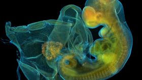
But growing nervous tissue, such as spinal cords, in a dish is considerably trickier due to the complex structures and types of cell involved. During development, our embryos possess a structure called the neural tube, which is the precursor to the brain and spinal cord. This complex tissue begins to form during the third week after fertilization in a process called neurulation. The neural tube forms from a flat sheet of cells, called the neural plate, which undergoes a series of folds and deformations that ultimately result in a hollow tube. Because the neural tube has such a complex architecture, scientists have struggled to use scaffolds as a means to grow this tissue in the lab.
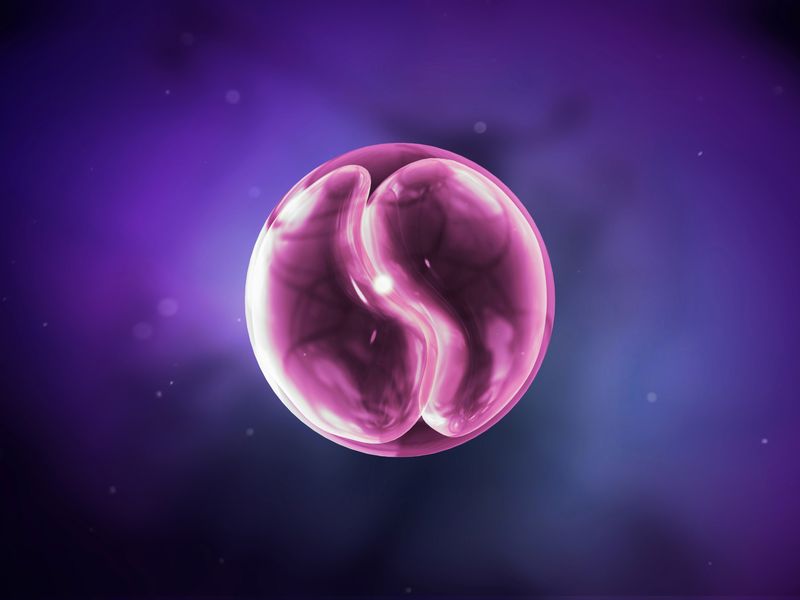
However, progress was made in the field when scientists discovered that embryonic stem cells grown in 3D suspension could self-organize into complex tissues when fed the right cocktail of ingredients. Building on this, researchers embedded single-cell suspensions of mouse embryonic stem cells within a 3D nutrient gel and added medium that is known to stimulate cells to differentiate into neural cells. When left untreated, the cells began to assume the identity of immature neurons, and produced small spherical structures called neuroepithelial cysts.
Next, they added a signaling molecule called retinoic acid which is known to lead to the activation of genes, such as sonic hedgehog, in the developing embryo. These are responsible for the differentiation of neural tube cells, or neural induction. They found that the cells began to self-organize in the dish, arranging themselves into the same patterns observed in the embryonic spinal cord. Using fluorescent antibodies, the researchers were able to confirm the presence of numerous different types of neuron, including motor neurons and relay neurons.
While the researchers have not tried out the technique using human stem cells, it raises the possibility that this could be achieved in the future, which could possibly serve as a source of tissue for patients with spinal cord injuries.
[Via Stem Cell Reports and The Guardian]