

Mammals are pretty complex things, made up of a huge number of perfectly specialized cells that perform a massive range of vital functions. What’s even more amazing is that all start out as just a single zygote – or fertilized egg – that then divides in order to form an embryo. The moment at which the dividing cells first become differentiated from one another has now been caught on film, enabling scientists to identify the precise point in early mammalian development at which this occurs.
The rest of this article is behind a paywall. Please sign in or subscribe to access the full content.Previously, researchers had not been able to capture this initial stage of cell division on camera, since the cells involved in this process are extremely light-sensitive. However, scientists at the European Molecular Biology Laboratory have finally managed to achieve the feat, using a technique called light-sheet microscopy, which illuminates only a tiny strip of a sample in order to minimize exposure.
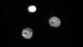
Above is the video of the specialization. Credit: European Molecular Biology Laboratory
Publishing their findings in the journal Nature Methods, the team explain how they recorded the early development of 139 mouse embryos, focusing particularly on the stage at which the zygote becomes a blastocyst. This is a small bundle of 64 cells, made up of an outer layer called the trophectoderm (TE), which goes on to form the placenta, and an inner cell mass (ICM), which later develops into the foetus. This division marks the first moment of cellular differentiation, which is why the researchers were so keen to locate the exact moment at which this occurs.
Observing the process in action, they found that this differentiation of dividing cells occurs when the embryo expands from 8 to 16 cells. As can be seen in the footage, it is at this stage the cells begin to behave in a specialized manner, with the ICM – marked in red in the video – grouping together at the center of the bundle while the cells of the TE arrange themselves around the outside.
Mouse blastocysts are made up of a smaller number of cells than human blastocysts, so it does not necessarily follow that cell differentiation occurs at the same stage in human embryonic development. However, the fact that it is now possible to observe the progression of mammalian zygotes to blastocysts opens up new possibilities for mapping the action of molecules that drive this process. This, according to the study authors, could have a number of implications for the treatment of human infertility.