

No magic wands necessary: Scientists have managed to levitate cells in the lab in a bid to improve disease diagnostics. By tweaking a technique used in space science to detect tiny faults in rockets, the scientists could manipulate cells with sound waves and determine their “stiffness,” which is a tell-tale sign of certain diseases.
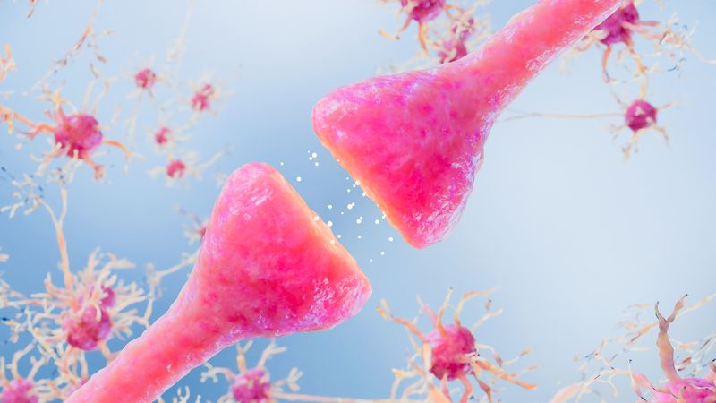
Cells maintain their shape with a structural framework known as the cytoskeleton, which consists of a scaffold-like network of filaments and microscopic tubes. While the mechanical properties of cells are altered in many diseases, there are some in which cytoskeletal changes are a hallmark characteristic, like cancer and some neurodegenerative diseases.
“During the metastasis of cancer, for example, the cell’s cytoskeleton becomes more elastic to allow the cells to migrate through tissue,” researcher Brian Patchett from Utah Valley University explained to IFLScience. “Similarly, it has been found that more aggressive breast cancers have a different stiffness than less aggressive types.”
But why the need for the Harry Potter style trick? Prior attempts by the team to assess this property in cells grown the traditional way – in petri dishes – failed because reflections from the surface interfered with measurements, Patchett said. Furthermore, the shape of cells grown in this manner isn’t an accurate representation of their native state in the body, as they get squished against the dish.
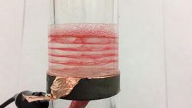
So what the team did was develop a technique that allowed them to acoustically suspend cells in layers in a fluid, enabling them to take measurements with minimal interference. This involved the use of two types of ultrasonic waves: low-frequency for the wizardry, and high-frequency to assess stiffness. For the first trick, they had to create something known as a standing wave, which involved trapping soundwaves between a transducer (something that converts energy from one form to another) and a reflective surface.
Standing waves, as the name suggests, don’t travel; they still vibrate, but they don’t propagate like traveling waves. Although they don’t go anywhere, they still have areas of high disturbance, called antinodes, and regions that are a lot calmer, nodes, where pressure stays pretty much the same. It is in the latter where the cells collect in layers. After performing this feat, the scientists measured the cells’ properties by probing their innards using high-frequency ultrasound.
To offer superiority over current methods, the method has got to offer fast results, especially if doctors want to use it in the operating room to assess cancer aggressiveness, as the researchers claim it could. Patchett said that their current method offers pathology results in around three minutes for cell samples, but solid tissue takes about 20 minutes longer because the cells need to be prepared in a solution first. But that’s still an improvement over some histology techniques, which can take days.
Ultimately, the goal is to develop this technique further so that it could be used in the emerging field of personalized medicine, whereby treatments are tailored to the individual patient’s needs, rather than using one-size-fits-all treatments that can have low efficacy rates and risk side effects. The work, currently unpublished but presented at the Acoustical Society of America's fall 2015 meeting, clearly has some way to go before it reaches the clinic, but if the technique could offer a quick turnover of reliable results, it would be a welcomed addition.