

Researchers have developed stretchy, bendy electronics that are so thin they can be rolled up and jammed into a small needle with a 0.1-millimeter diameter, then injected into living tissue. Within an hour of being injected into the brains of live mice, the electronics unfurled and began monitoring biological activity. The work is described in Nature Nanotechnology this week.
Flexible, implantable electronics would make it possible for continuous biomonitoring from the surface of non-flat structures like internal cavities. These would have a host of useful biomedical applications, such as checking electrophysiological signals related to epilepsy and arrhythmia. Previous research revealed that electronics like these can be surgically implanted, but so far, it hasn’t been possible to precisely control their delivery to target areas within the body non-invasively.

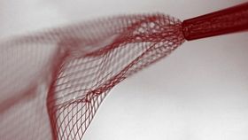
Now, a team led by Harvard’s Charles Lieber and Ying Fang from the National Center for Nanoscience and Technology in Beijing has designed syringe-injectable, mesh-shaped electronics (top image) consisting of a polymer–metal combination. Once rolled up and loaded into a syringe – which can have a diameter as small as 100 micrometers – the electrical components can be injected into cavities or specific regions of living tissues. After the jab, as the needle is withdrawn, the electronics unfold to about 80% of their original configuration – with no loss of function, the team reports. The photo directly above shows the injection of mesh electronics through a metal needle into an aqueous solution. Mesh electronics with widths more than 30 times that of the metal or glass needle can be successfully injected.
 To examine the behavior of the mesh electronics, the team used a glass needle to inject them into anaesthetized mice in two distinct brain regions: the lateral ventricle and the hippocampus. Over a five-week period, the mice showed no immune response, and the foldable electrical units even began to network with the healthy neurons. To the right is a 3D microscopy image of mesh electronics that have been injected into the lateral ventricle. You can see the innervation of the neural tissue, as well as the migration of neural progenitor cells onto the mesh within the cavity.
Furthermore, the team was able to monitor brain activity using the electronics (and with input and output wiring) in the hippocampus with limited damage to the surrounding tissue.
Images: Lieber Research Group, Harvard University