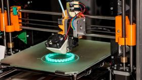


3D printing is one of a number of technologies that have bloomed and spread across the world incredibly rapidly in the last decade. Prosthetics for amputees, rocket parts, and bridges have all been built using 3D printers, and recently, researchers demonstrated how to print out the structures for human organs using biological material. This month, a new study in the journal Biofabrication has described the process of 3D printing stem cells' “building blocks” for the first time.
Broadly, there are two main types of stem cells: adult, which are located in a range of biological tissue types, and embryonic (ESCs), which exist in the cell mass within animals that subsequently develops into an embryo. This new technique – called bioprinting – is able to produce the structural “building blocks” of the latter in 3D. Although 3D sheets of cells have been produced before, this novel technique generates 3D structures that are optimized for the growth of ESCs.
Routinely, during development, ESCs clump together into spherical “embryoid bodies” (EBs), gatherings of pluripotent cells that then go on to develop into any cell type in the body. Although researchers can grow ESCs on flat plates or sheets, a 3D spherical structure is more apt for this type of growth, which the team hoped to replicate.
A suspension of ESCs was mixed with hydrogel, a “smart” material that changes its structure in response to acidity, temperature and other variables. Using this “bioink,” a six-layer grid square 8 millimeters (0.32 inches) across and 1 millimeter (0.04 inches) thick was printed, and enticed to grow. The cells developed into spherical EBs without leaving their individual grid sections, meaning that they divided and multiplied without clumping randomly across the grid.
The result was a uniform production of EBs across the entire grid, in a process mimicking embryo formation, growing far faster than they would have done on a flat surface. The researchers note that it is very easy to damage stem cells during the bioprinting process; remarkably, 90% of their printed ESCs remained fully functional and capable of self-renewing seven days after printing. In addition, their ability to differentiate into other types of cells remained unaffected a week on from their creation.
ESCs can be grown in the lab using a variety of methods, but exercising control on their growth and development is notoriously difficult. Growing their ESCs in 3D, this team of researchers were able to produce genuine EBs with an unprecedentedly high degree of precision.
Wei Sun, the lead author of the study, said in a statement: “I think that we've produced a 3-D microenvironment which is much more like that found in [the human body] for growing embryoid bodies, which explains the higher levels of cell proliferation (growth).
“In the longer term, we'd like to produce controlled heterogeneous (different types of) embryonic bodies,” Sun continued. “This would promote different cell types developing next to each other - which would lead the way for growing micro-organs from scratch within the lab.”