

Researchers have developed a microscopy technique that is 6.6 times more sensitive than other current techniques of its type, as shown in a paper published in Light: Science and Applications. The new method could even be powerful enough to observe things as small as a virus. “Small signals from nanoscale particles like viruses or particles moving around inside and outside a cell could be detected, which allows for simultaneous observation of their behavior and the cell's state," said the first author of the study Takuro Ideguchi, Associate Professor at the University of Tokyo Institute for Photon Science and Technology, in a statement.
The rest of this article is behind a paywall. Please sign in or subscribe to access the full content.The new technique is called adaptive dynamic range shift quantitative phase imaging (ADRIFT-QPI) and is an upgrade to the existing quantitative phase imaging method. Quantitative phase imaging is already powerful enough to clearly observe the roughness of a glass slide and is commonly used to observe biological processes. It works by sending a flat sheet of light in a pulse towards the sample and then measuring the phase shift, which is how different the light is when it emerges on the other side.
Professor Ideguchi stated that "To see greater detail using the same image sensor, we must expand the dynamic range so that we can detect smaller phase changes of light," and the University of Tokyo team saw a way to achieve this.
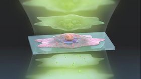
ADRIFT-QPI improves sensitivity by using a two-exposure method. The first exposure pulses the sample with a flat sheet of light as usual. Computer analysis then designs a “sculpture of light” based on the reading from the first pulse. This sculpted wavefront is pulsed at the sample at a higher light intensity than the first pulse. This second pulse reveals minuscule details that would be drowned out in the first pulse. A program then stitches these two readings together, forming the final image.
Professor Ideguchi also explains that "Our ADRIFT-QPI method needs no special laser, no special microscope or image sensors; we can use live cells, we don't need any stains or fluorescence, and there is very little chance of phototoxicity." The illumination intensity of ADRIFT-QPI is lower than in other imaging techniques used on live cells. This reduces damage to cells via light, known as phototoxicity. The method not requiring stains or fluorescent dyes is significant, as these can be expensive, hard to use, and potentially affect the molecule they are attached to. Not requiring these dyes, as well as not needing to buy more special equipment, could save future researchers time, effort, and money. This, combined with impressive imaging capabilities, makes ADRIFT-QPI a very promising microscopy method.