

Ladies and gentlemen, meet the skinbow zebrafish. This genetically modified aquatic creature has technicolor scales, meaning that it can broadcast all the colors of the rainbow to any passing observer.
Far from being a marvel of synthetic biology, however, this beautiful little freshwater critter does serve a more practical use. As the new study in the journal Developmental Cell showcases, this zebrafish will help researchers examine the way tissue regenerates on a cellular level, something that is notoriously difficult to track due to its complex nature.
Zebrafish have remarkable tissue regeneration skills, including the ability to heal damaged limbs, hearts and even their spinal cords. Consequently, researchers are fond of using them to study how cells divide, form and repair different parts of their anatomy.
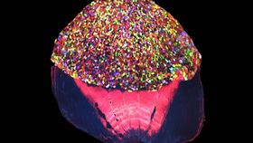
By genetically modifying the DNA within its skin cells in order to contain red, blue and green fluorescent “tags,” the researchers have given their zebrafish the ability to express its scales using 5,000 different hues – although only 70 are able to be differentiated under a microscope. Not limited to just having fluorescing, colorful scales, this particular fish is even able to exhibit intricate color patterns within the thin, translucent cornea shielding its own eyes.
This remarkable iridescence has earned it the moniker of “skinbow.” This technique is an offshoot of the “brainbow” concept: In 2007, a study revealed that by genetically altering the DNA within neurons, they were able to exhibit a range of fluorescent proteins, producing a plethora of colors.
In the case of the skinbow, the unique color “fingerprint” of each of the individually colored zebrafish cells meant that each and every one of them could be carefully tracked by researchers. “It is like you have given each cell an individual barcode,” said Chen-Hui Chen, a postdoctoral fellow at Duke University and lead author on the study, in a statement.
The unsuspecting, shimmering zebrafish was then subjected to several flesh wounds, some harsher than others. The point was to see how the skin cells responded to the injuries, and to the researchers’ delight, they could observe this in real-time with unprecedented precision.

Every cell shows up as a color combination of red, blue and green. Chen et al./Developmental Cell
During the amputation of a fin, they saw how skin cells migrated from nearby areas to rapidly conceal the incision; then, new skin cells were manufactured in order to reinforce the recruited, older cells. Finally, the skin cells expanded in size in order to cover more of the wound, allowing it to properly heal.
Far from just observing how injuries naturally heal, the skinbow could also be used in future research to show how various drugs slow down or speed up tissue regeneration. In addition, various infections or diseases, including cancer, could be inflicted on the poor genetically modified zebrafish, and the subsequent movement of the skinbow cells will help researchers track how they respond to the threat.
“Before we can fully understand tissue regeneration, we need to be able to monitor what individual cells are doing,” said Kenneth Poss, a professor of cell biology at Duke University and one of the authors of the study. “This is a cutting-edge way to visualize hundreds or thousands of cells at once in a regenerating tissue.”