

Ultrasound scans are very useful for peering inside the body; using high-frequency sound waves, doctors can create images of various organs to detect problems, or monitor developing babies in the womb. But it turns out that ultrasound scans may have another, rather unexpected use in medicine: They may help treat Alzheimer’s disease.
The rest of this article is behind a paywall. Please sign in or subscribe to access the full content.In a new study, scientists found that scanning the brains of mice with a model of this disease not only helped clear the abnormal protein buildups that are associated with Alzheimer’s, but it also improved their memory. Although mice are very different from us, if it turns out to be safe and effective in humans, it could be an invaluable tool not only for the treatment of Alzheimer’s, but other brain diseases as well. The study has been published in Science Translational Medicine.
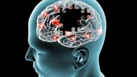
Alzheimer’s disease (AD) is a neurodegenerative disorder characterized by progressive cognitive impairment. Although the cause of AD remains to be elucidated, we know that its development is associated with brain shrinkage and the formation of characteristic plaques in certain areas. These consist of dead cells and a protein known as amyloid-beta (Aβ), which aggregates into abnormal fibers that accumulate as toxic clumps outside of cells.
Much research into AD has therefore focused on preventing the buildup of these aggregates or clearing them from the brain, but scientists are faced with a problem: The brain is shielded by a delicate layer of cells, known as the blood-brain barrier (BBB), which is very difficult for things to cross, such as therapeutics or helpful antibodies. Scientists have therefore been endeavoring to develop ways to temporarily open it up, allowing drugs or components of the immune system to get inside, which could help clear the problematic proteins.
A few years back, scientists found that combining ultrasound with harmless microscopic bubbles, which are used as ultrasound contrast agents, could be an effective technique. The sound waves cause the bubbles to vibrate and expand, which creates transient openings in the BBB. Researchers therefore wondered whether this technique could boost our brain’s protective response to the toxic Aβ proteins. Our brain is equipped with cells, called microglia, which usually mop-up harmful proteins like this, but in AD proteins these are impaired. By allowing components of the immune system through the BBB, they could act to stimulate these cells back into action.
To test this hypothesis, scientists from the Queensland Brain Institute used mice with models of AD and injected them with microbubbles before repeatedly scanning the entire brains of half the animals, leaving the others as a control. They found that the animals receiving ultrasound showed activation of microglia in their brains, which had extensively engulfed Aβ proteins. Compared to the controls, the Aβ burden was also reduced as 75% of the mice had cleared almost all of their plaques. Furthermore, these animals also performed better on three different memory tasks.
While this is certainly promising, we don’t know whether it would be safe or effective in humans. Some experts have pointed out that although the treatment is not invasive, it is still disruptive and the potential negative consequences of letting our brain’s guard down, even temporarily, are not made clear by this study. Further research is therefore warranted, so the researchers plan to use larger animals next.